 Movie
Movie Controller

[English] 日本語
 Yorodumi
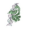
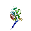
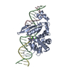
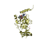
- PDB-1mjh: Structure-based assignment of the biochemical function of hypothe... -
+ Open data

- Basic information
| Entry | Database: PDB / ID: 1mjh | ||||||
|---|---|---|---|---|---|---|---|
| Title | Structure-based assignment of the biochemical function of hypothetical protein MJ0577: A test case of structural genomics | ||||||
 Components Components | PROTEIN (ATP-BINDING DOMAIN OF PROTEIN MJ0577) | ||||||
 Keywords Keywords | HYPOTHETICAL PROTEIN / STRUCTURAL GENOMICS / FUNCTIONAL ASSIGNMENT / ATP BINDING PROTEIN / BSGC structure funded by NIH / Protein Structure Initiative / PSI / Berkeley Structural Genomics Center | ||||||
| Function / homology |  Function and homology information Function and homology information | ||||||
| Biological species |   Methanocaldococcus jannaschii (archaea) Methanocaldococcus jannaschii (archaea) | ||||||
| Method |  X-RAY DIFFRACTION / SYNCHROTRON / MAD / Resolution: 1.7 Å X-RAY DIFFRACTION / SYNCHROTRON / MAD / Resolution: 1.7 Å | ||||||
 Authors Authors | Zarembinski, T.I. / Hung, L.-W. / Mueller-Dieckmann, H.J. / Kim, K.-K. / Yokota, H. / Kim, R. / Kim, S.-H. / Berkeley Structural Genomics Center (BSGC) | ||||||
 Citation Citation | Journal: Proc.Natl.Acad.Sci.USA / Year: 1998 Title: Structure-based assignment of the biochemical function of a hypothetical protein: a test case of structural genomics. Authors: Zarembinski, T.I. / Hung, L.-W. / Mueller-Dieckmann, H.J. / Kim, K.-K. / Yokota, H. / Kim, R. / Kim, S.-H. | ||||||
| History |
|
- Structure visualization
| Structure viewer | Molecule: MolmilJmol/JSmol |
|---|
- Downloads & links
-Download
| PDBx/mmCIF format | 1mjh.cif.gz | 79 KB | Display | PDBx/mmCIF format |
|---|---|---|---|---|
| PDB format | pdb1mjh.ent.gz | 58.5 KB | Display | PDB format |
| PDBx/mmJSON format | 1mjh.json.gz | Tree view | PDBx/mmJSON format | |
| Others |  Other downloads Other downloads |
-Validation report
| Arichive directory | https://data.pdbj.org/pub/pdb/validation_reports/mj/1mjhftp://data.pdbj.org/pub/pdb/validation_reports/mj/1mjh | HTTPS FTP |
|---|
-Related structure data
| Similar structure data | |
|---|---|
| Other databases |


-Links
 PDBj
PDBj- Assembly
| Deposited unit | 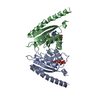
| ||||||||||
|---|---|---|---|---|---|---|---|---|---|---|---|
| 1 |
| ||||||||||
| Unit cell |
|
-Components
| #1: Protein | Mass: 18370.600 Da / Num. of mol.: 2 / Fragment: ATP-BINDING DOMAIN Source method: isolated from a genetically manipulated source Details: COMPLEXED WITH ADENOSINE-5'-TRIPHOSPHATE Source: (gene. exp.) Methanocaldococcus jannaschii (archaea)Description: RECENTLY SEQUENCED HYPERTHERMOPHILE / Cellular location: CYTOPLASM / Gene: MJ0577 / Plasmid: PET-23A / Production host:  #2: Chemical |   Mass: 54.938 Da / Num. of mol.: 2 / Source method: obtained synthetically / Formula: Mn Mass: 54.938 Da / Num. of mol.: 2 / Source method: obtained synthetically / Formula: Mn#3: Chemical |   Mass: 507.181 Da / Num. of mol.: 2 / Source method: obtained synthetically / Formula: C10H16N5O13P3 / Comment: ATP, energy-carrying molecule*YM Mass: 507.181 Da / Num. of mol.: 2 / Source method: obtained synthetically / Formula: C10H16N5O13P3 / Comment: ATP, energy-carrying molecule*YM#4: Water | ChemComp-HOH / |  Mass: 18.015 Da / Num. of mol.: 284 / Source method: isolated from a natural source / Formula: H2O Mass: 18.015 Da / Num. of mol.: 284 / Source method: isolated from a natural source / Formula: H2O |
|---|
-Experimental details
-Experiment
| Experiment | Method: X-RAY DIFFRACTION / Number of used crystals: 1 |
|---|
- Sample preparation
| Crystal | Density Matthews: 2.34 Å3/Da / Density % sol: 47.48 % | ||||||||||||||||||||||||||||||||||||||||||||||||||||||||||||
|---|---|---|---|---|---|---|---|---|---|---|---|---|---|---|---|---|---|---|---|---|---|---|---|---|---|---|---|---|---|---|---|---|---|---|---|---|---|---|---|---|---|---|---|---|---|---|---|---|---|---|---|---|---|---|---|---|---|---|---|---|---|
| Crystal grow | Method: vapor diffusion, hanging drop / pH: 7 / Details: pH 7.0, VAPOR DIFFUSION, HANGING DROP | ||||||||||||||||||||||||||||||||||||||||||||||||||||||||||||
| Crystal grow | *PLUS Temperature: 100 K | ||||||||||||||||||||||||||||||||||||||||||||||||||||||||||||
| Components of the solutions | *PLUS
|
-Data collection
| Diffraction | Mean temperature: 100 K | |||||||||||||||
|---|---|---|---|---|---|---|---|---|---|---|---|---|---|---|---|---|
| Diffraction source | Source: SYNCHROTRON / Site: ALS  / Type: ALS / Wavelength: 0.9683, 0.9799, 0.9806, 1.000 / Type: ALS / Wavelength: 0.9683, 0.9799, 0.9806, 1.000 | |||||||||||||||
| Detector | Detector: CCD / Date: Mar 15, 1998 | |||||||||||||||
| Radiation | Protocol: MAD / Monochromatic (M) / Laue (L): M / Scattering type: x-ray | |||||||||||||||
| Radiation wavelength |
| |||||||||||||||
| Reflection | Resolution: 1.7→20 Å / Num. obs: 38706 / % possible obs: 98 % / Redundancy: 7.5 % / Rsym value: 0.049 | |||||||||||||||
| Reflection shell | Highest resolution: 1.7 Å / Rsym value: 0.0249 / % possible all: 85.4 | |||||||||||||||
| Reflection | *PLUS Num. measured all: 292156 / Rmerge(I) obs: 0.049 | |||||||||||||||
| Reflection shell | *PLUS % possible obs: 85.4 % / Rmerge(I) obs: 0.249 |
- Processing
| Software |
| |||||||||||||||||||||||||||||||||||||||||||||||||||||||||||||||
|---|---|---|---|---|---|---|---|---|---|---|---|---|---|---|---|---|---|---|---|---|---|---|---|---|---|---|---|---|---|---|---|---|---|---|---|---|---|---|---|---|---|---|---|---|---|---|---|---|---|---|---|---|---|---|---|---|---|---|---|---|---|---|---|---|
| Refinement | Method to determine structure: MAD / Resolution: 1.7→20 Å / Cross valid method: THROUGHOUT / σ(F): 0 Details: REFINEMENT WAS BEGUN WITH CNS AND FINAL REFINEMENT WITH REFMAC AND ARP
| |||||||||||||||||||||||||||||||||||||||||||||||||||||||||||||||
| Displacement parameters | Biso mean: 30.1 Å2 | |||||||||||||||||||||||||||||||||||||||||||||||||||||||||||||||
| Refinement step | Cycle: LAST / Resolution: 1.7→20 Å
| |||||||||||||||||||||||||||||||||||||||||||||||||||||||||||||||
| Refine LS restraints |
| |||||||||||||||||||||||||||||||||||||||||||||||||||||||||||||||
| Software | *PLUS Name: REFMAC / Classification: refinement | |||||||||||||||||||||||||||||||||||||||||||||||||||||||||||||||
| Refinement | *PLUS Rfactor obs: 0.21 | |||||||||||||||||||||||||||||||||||||||||||||||||||||||||||||||
| Solvent computation | *PLUS | |||||||||||||||||||||||||||||||||||||||||||||||||||||||||||||||
| Displacement parameters | *PLUS |
