 Movie
Movie Controller


+ Open data

- Basic information
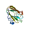
| Entry | Database: PDB / ID: 1mb1 | ||||||
|---|---|---|---|---|---|---|---|
| Title | MBP1 FROM SACCHAROMYCES CEREVISIAE | ||||||
 Components Components | MLU1-BOX BINDING PROTEIN | ||||||
 Keywords Keywords | TRANSCRIPTION REGULATION / CELL-CYCLE / TRANSCRIPTION FACTOR | ||||||
| Function / homology |  Function and homology information Function and homology informationMBF transcription complex / SBF transcription complex / G1/S transition of mitotic cell cycle / maintenance of translational fidelity / DNA-binding transcription activator activity, RNA polymerase II-specific / sequence-specific DNA binding / regulation of DNA-templated transcription / positive regulation of transcription by RNA polymerase II / nucleus Similarity search - Function | ||||||
| Biological species |  | ||||||
| Method |  X-RAY DIFFRACTION / SYNCHROTRON / MAD / Resolution: 2.1 Å X-RAY DIFFRACTION / SYNCHROTRON / MAD / Resolution: 2.1 Å | ||||||
 Authors Authors | Taylor, I.A. / Smerdon, S.J. | ||||||
 Citation Citation | Journal: J.Mol.Biol. / Year: 1997 Title: The X-ray structure of the DNA-binding domain from the Saccharomyces cerevisiae cell-cycle transcription factor Mbp1 at 2.1 A resolution. Authors: Taylor, I.A. / Treiber, M.K. / Olivi, L. / Smerdon, S.J. | ||||||
| History |
|
- Structure visualization
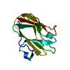
| Structure viewer | Molecule: MolmilJmol/JSmol |
|---|
- Downloads & links
-Download
| PDBx/mmCIF format | 1mb1.cif.gz | 33.6 KB | Display | PDBx/mmCIF format |
|---|---|---|---|---|
| PDB format | pdb1mb1.ent.gz | 22.3 KB | Display | PDB format |
| PDBx/mmJSON format | 1mb1.json.gz | Tree view | PDBx/mmJSON format | |
| Others |  Other downloads Other downloads |
-Validation report
| Arichive directory | https://data.pdbj.org/pub/pdb/validation_reports/mb/1mb1ftp://data.pdbj.org/pub/pdb/validation_reports/mb/1mb1 | HTTPS FTP |
|---|
-Related structure data
| Similar structure data |
|---|


-Links
 PDBj
PDBj

- Assembly
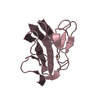
| Deposited unit | 
| ||||||||
|---|---|---|---|---|---|---|---|---|---|
| 1 |
| ||||||||
| Unit cell |
|
-Components
| #1: Protein | Mass: 14900.885 Da / Num. of mol.: 1 / Fragment: DNA-BINDING DOMAIN Source method: isolated from a genetically manipulated source Source: (gene. exp.) Cell line: BL21 / Cellular location: NUCLEAR / Plasmid: PET22B / Species (production host): Escherichia coli / Production host:  |
|---|---|
| #2: Water | ChemComp-HOH /  Mass: 18.015 Da / Num. of mol.: 60 / Source method: isolated from a natural source / Formula: H2O Mass: 18.015 Da / Num. of mol.: 60 / Source method: isolated from a natural source / Formula: H2O |
-Experimental details
-Experiment
| Experiment | Method: X-RAY DIFFRACTION / Number of used crystals: 1 |
|---|
- Sample preparation
| Crystal | Density Matthews: 1.88 Å3/Da / Density % sol: 28 % | ||||||||||||||||||||||||
|---|---|---|---|---|---|---|---|---|---|---|---|---|---|---|---|---|---|---|---|---|---|---|---|---|---|
| Crystal grow | pH: 6.9 / Details: pH 6.9 | ||||||||||||||||||||||||
| Crystal | *PLUS | ||||||||||||||||||||||||
| Crystal grow | *PLUS Temperature: 4 ℃ / Method: vapor diffusion, hanging dropDetails: Taylor, I.A., (1997) Proteins Struct. Funct. Genet., 27,325. PH range low: 8.2 / PH range high: 7.7 | ||||||||||||||||||||||||
| Components of the solutions | *PLUS
|
-Data collection
| Diffraction | Mean temperature: 100 K |
|---|---|
| Diffraction source | Source: SYNCHROTRON / Site: ELETTRA  / Beamline: 5.2R / Wavelength: 1.22 / Beamline: 5.2R / Wavelength: 1.22 |
| Detector | Type: MARRESEARCH / Detector: IMAGE PLATE AREA DETECTOR / Date: Jan 1, 1997 |
| Radiation | Monochromatic (M) / Laue (L): M / Scattering type: x-ray |
| Radiation wavelength | Wavelength: 1.22 Å / Relative weight: 1 |
| Reflection | Resolution: 2.1→18 Å / Num. obs: 6810 / % possible obs: 94.4 % / Redundancy: 4.7 % / Biso Wilson estimate: 32 Å2 / Rmerge(I) obs: 0.041 / Net I/σ(I): 30 |
| Reflection shell | Resolution: 2.1→2.16 Å / Rmerge(I) obs: 0.124 / Mean I/σ(I) obs: 11 / % possible all: 93.3 |
- Processing
| Software |
| |||||||||||||||||||||||||||||||||||||||||||||||||||||||||||||||
|---|---|---|---|---|---|---|---|---|---|---|---|---|---|---|---|---|---|---|---|---|---|---|---|---|---|---|---|---|---|---|---|---|---|---|---|---|---|---|---|---|---|---|---|---|---|---|---|---|---|---|---|---|---|---|---|---|---|---|---|---|---|---|---|---|
| Refinement | Method to determine structure: MAD / Resolution: 2.1→7 Å / Cross valid method: THROUGHOUT
| |||||||||||||||||||||||||||||||||||||||||||||||||||||||||||||||
| Displacement parameters | Biso mean: 27 Å2 | |||||||||||||||||||||||||||||||||||||||||||||||||||||||||||||||
| Refinement step | Cycle: LAST / Resolution: 2.1→7 Å
| |||||||||||||||||||||||||||||||||||||||||||||||||||||||||||||||
| Refine LS restraints |
| |||||||||||||||||||||||||||||||||||||||||||||||||||||||||||||||
| Software | *PLUS Name: CCP4 / Classification: refinement | |||||||||||||||||||||||||||||||||||||||||||||||||||||||||||||||
| Refinement | *PLUS Rfactor obs: 0.215 | |||||||||||||||||||||||||||||||||||||||||||||||||||||||||||||||
| Solvent computation | *PLUS | |||||||||||||||||||||||||||||||||||||||||||||||||||||||||||||||
| Displacement parameters | *PLUS | |||||||||||||||||||||||||||||||||||||||||||||||||||||||||||||||
| Refine LS restraints | *PLUS Type: p_angle_deg / Dev ideal: 2.47 |
