 Movie
Movie Controller

[English] 日本語
 Yorodumi
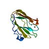
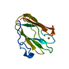
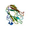
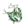
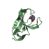
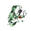
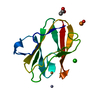
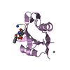
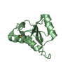
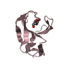
- PDB-1id2: CRYSTAL STRUCTURE OF AMICYANIN FROM PARACOCCUS VERSUTUS (THIOBACI... -
+ Open data

- Basic information
| Entry | Database: PDB / ID: 1id2 | ||||||
|---|---|---|---|---|---|---|---|
| Title | CRYSTAL STRUCTURE OF AMICYANIN FROM PARACOCCUS VERSUTUS (THIOBACILLUS VERSUTUS) | ||||||
 Components Components | AMICYANIN | ||||||
 Keywords Keywords | ELECTRON TRANSPORT / Beta Barrel / type-1 blue copper protein / electron transfer protein | ||||||
| Function / homology |  Function and homology information Function and homology information | ||||||
| Biological species |  Paracoccus versutus (bacteria) Paracoccus versutus (bacteria) | ||||||
| Method |  X-RAY DIFFRACTION / MOLECULAR REPLACEMENT / Resolution: 2.15 Å X-RAY DIFFRACTION / MOLECULAR REPLACEMENT / Resolution: 2.15 Å | ||||||
 Authors Authors | Romero, A. / Nar, H. / Messerschmidt, A. | ||||||
 Citation Citation | Journal: J.Mol.Biol. / Year: 1994 Title: Crystal structure analysis and refinement at 2.15 A resolution of amicyanin, a type I blue copper protein, from Thiobacillus versutus. Authors: Romero, A. / Nar, H. / Huber, R. / Messerschmidt, A. / Kalverda, A.P. / Canters, G.W. / Durley, R. / Mathews, F.S. | ||||||
| History |
|
- Structure visualization
| Structure viewer | Molecule: MolmilJmol/JSmol |
|---|
- Downloads & links
-Download
| PDBx/mmCIF format | 1id2.cif.gz | 76.1 KB | Display | PDBx/mmCIF format |
|---|---|---|---|---|
| PDB format | pdb1id2.ent.gz | 57.3 KB | Display | PDB format |
| PDBx/mmJSON format | 1id2.json.gz | Tree view | PDBx/mmJSON format | |
| Others |  Other downloads Other downloads |
-Validation report
| Arichive directory | https://data.pdbj.org/pub/pdb/validation_reports/id/1id2ftp://data.pdbj.org/pub/pdb/validation_reports/id/1id2 | HTTPS FTP |
|---|
-Related structure data
| Related structure data |  1aanS S: Starting model for refinement |
|---|---|
| Similar structure data |


-Links
 PDBj
PDBj- Assembly
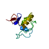
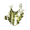
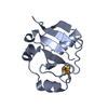
| Deposited unit | 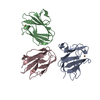
| ||||||||
|---|---|---|---|---|---|---|---|---|---|
| 1 | 
| ||||||||
| 2 | 
| ||||||||
| 3 | 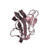
| ||||||||
| Unit cell |
| ||||||||
| Details | The three independent molecules of amicyanin are arranged as columns around 3(2) axes centered at (0, 0) molecule C, (1/3, 2/3) molecule B and (2/3, 1/3) molecule A. The following transformation relate molecule A to B. (-0.975 -0.205 0.081) 49.109 (-0.208 0.977 -0.044) 33.553 (-0.070 -0.060 -0.996) -13.650 A to C: (0.240 -0.970 0.033) 14.988 (-0.969 -0.238 0.052) 49.233 -0.043 -0.044 -0.998) -16.988 C to B: (-0.034 0.999 -0.028) -0.062 (-0.999 -0.034 0.012) 50.335 (0.011 0.028 0.999) 1.745 |
-Components
| #1: Protein | Mass: 11703.384 Da / Num. of mol.: 3 Source method: isolated from a genetically manipulated source Source: (gene. exp.) Paracoccus versutus (bacteria) / Gene: MAUC OR AMI / Plasmid: PUC18 / Production host: #2: Chemical |   Mass: 63.546 Da / Num. of mol.: 3 / Source method: obtained synthetically / Formula: Cu Mass: 63.546 Da / Num. of mol.: 3 / Source method: obtained synthetically / Formula: Cu#3: Water | ChemComp-HOH / |  Mass: 18.015 Da / Num. of mol.: 122 / Source method: isolated from a natural source / Formula: H2O Mass: 18.015 Da / Num. of mol.: 122 / Source method: isolated from a natural source / Formula: H2OHas protein modification | N | |
|---|
-Experimental details
-Experiment
| Experiment | Method: X-RAY DIFFRACTION / Number of used crystals: 1 |
|---|
- Sample preparation
| Crystal | Density Matthews: 2.4 Å3/Da / Density % sol: 48.71 % | ||||||||||||||||||||||||||||||||||||||||||
|---|---|---|---|---|---|---|---|---|---|---|---|---|---|---|---|---|---|---|---|---|---|---|---|---|---|---|---|---|---|---|---|---|---|---|---|---|---|---|---|---|---|---|---|
| Crystal grow | Temperature: 293 K / Method: vapor diffusion, hanging drop / pH: 4.5 Details: Reservoir: 2.05 M ammonium sulfate, 0.1 M Na, K phosphate; hanging drop: 5 microliter protein solution (6 mg/ ml) plus 4 microliter 10% (v/v) of MPD and 1.5 microliter 0.5% (v/v) beta-D- ...Details: Reservoir: 2.05 M ammonium sulfate, 0.1 M Na, K phosphate; hanging drop: 5 microliter protein solution (6 mg/ ml) plus 4 microliter 10% (v/v) of MPD and 1.5 microliter 0.5% (v/v) beta-D-glucopyranoside, pH 4.5, VAPOR DIFFUSION, HANGING DROP, temperature 293K | ||||||||||||||||||||||||||||||||||||||||||
| Crystal grow | *PLUS PH range low: 5 / PH range high: 4 | ||||||||||||||||||||||||||||||||||||||||||
| Components of the solutions | *PLUS
|
-Data collection
| Diffraction | Mean temperature: 277 K |
|---|---|
| Diffraction source | Source: ROTATING ANODE / Type: RIGAKU RU200 / Wavelength: 1.5418 Å |
| Detector | Type: MARRESEARCH / Detector: IMAGE PLATE / Date: Jan 21, 1993 / Details: Ni-filter |
| Radiation | Monochromator: Ni-Filter / Protocol: SINGLE WAVELENGTH / Monochromatic (M) / Laue (L): M / Scattering type: x-ray |
| Radiation wavelength | Wavelength: 1.5418 Å / Relative weight: 1 |
| Reflection | Resolution: 2.15→25 Å / Num. all: 92670 / Num. obs: 84144 / % possible obs: 90.8 % / Observed criterion σ(F): 0 / Observed criterion σ(I): 0 / Redundancy: 5.2 % / Rmerge(I) obs: 0.073 / Rsym value: 0.079 |
| Reflection shell | Resolution: 2.15→2.3 Å / Rmerge(I) obs: 0.345 / Rsym value: 0.383 / % possible all: 72.7 |
| Reflection | *PLUS Num. obs: 16083 / Num. measured all: 84144 |
| Reflection shell | *PLUS % possible obs: 72.7 % |
- Processing
| Software |
| |||||||||||||||||||||
|---|---|---|---|---|---|---|---|---|---|---|---|---|---|---|---|---|---|---|---|---|---|---|
| Refinement | Method to determine structure: MOLECULAR REPLACEMENT Starting model: PDB Entry 1AAN, these coordinates were supplied by the authors of this entry prior to their release Resolution: 2.15→8 Å / Isotropic thermal model: Isotropic / σ(F): 0 / σ(I): 0 / Stereochemistry target values: Engh & Huber
| |||||||||||||||||||||
| Displacement parameters | Biso mean: 24.6 Å2 | |||||||||||||||||||||
| Refine analyze | Luzzati coordinate error obs: 0.22 Å | |||||||||||||||||||||
| Refinement step | Cycle: LAST / Resolution: 2.15→8 Å
| |||||||||||||||||||||
| Refine LS restraints |
| |||||||||||||||||||||
| LS refinement shell | Resolution: 2.15→2.25 Å / Rfactor Rwork: 0.256 | |||||||||||||||||||||
| Software | *PLUS Name: X-PLOR / Version: 3.1 / Classification: refinement | |||||||||||||||||||||
| Refine LS restraints | *PLUS
|
