 Movie
Movie Controller

[English] 日本語
 Yorodumi
- PDB-1m04: Mutant Streptomyces plicatus beta-hexosaminidase (D313N) in compl... -
+ Open data

- Basic information
| Entry | Database: PDB / ID: 1m04 | ||||||
|---|---|---|---|---|---|---|---|
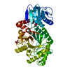
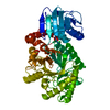
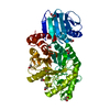
| Title | Mutant Streptomyces plicatus beta-hexosaminidase (D313N) in complex with product (GlcNAc) | ||||||
 Components Components | Beta-N-acetylhexosaminidase Hexosaminidase Hexosaminidase | ||||||
 Keywords Keywords | HYDROLASE / substrate assisted catalysis / hexosaminidase / family 20 glycosidase | ||||||
| Function / homology |  Function and homology informationbeta-N-acetylhexosaminidase / beta-N-acetylhexosaminidase activity / N-acetyl-beta-D-galactosaminidase activity / carbohydrate metabolic process Function and homology informationbeta-N-acetylhexosaminidase / beta-N-acetylhexosaminidase activity / N-acetyl-beta-D-galactosaminidase activity / carbohydrate metabolic processSimilarity search - Function | ||||||
| Biological species |  Streptomyces plicatus (bacteria) Streptomyces plicatus (bacteria) | ||||||
| Method | X-RAY DIFFRACTION / FOURIER SYNTHESIS / Resolution: 1.95 Å | ||||||
 Authors Authors | Williams, S.J. / Mark, B.L. / Vocadlo, D.J. / James, M.N.G. / Withers, S.G. | ||||||
 Citation Citation | Journal: J.Biol.Chem. / Year: 2002 Title: Aspartate 313 in the Streptomyces plicatus hexosaminidase plays a critical role in substrate-assisted catalysis by orienting the 2-acetamido group and stabilizing the transition state. Authors: Williams, S.J. / Mark, B.L. / Vocadlo, D.J. / James, M.N. / Withers, S.G. | ||||||
| History |
|
- Structure visualization
| Structure viewer | Molecule: MolmilJmol/JSmol |
|---|
- Downloads & links
-Download
| PDBx/mmCIF format | 1m04.cif.gz | 112.9 KB | Display | PDBx/mmCIF format |
|---|---|---|---|---|
| PDB format | pdb1m04.ent.gz | 90.2 KB | Display | PDB format |
| PDBx/mmJSON format | 1m04.json.gz | Tree view | PDBx/mmJSON format | |
| Others |  Other downloads Other downloads |
-Validation report
| Arichive directory | https://data.pdbj.org/pub/pdb/validation_reports/m0/1m04ftp://data.pdbj.org/pub/pdb/validation_reports/m0/1m04 | HTTPS FTP |
|---|
-Related structure data
| Related structure data |  1m01C 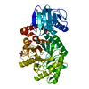 1m03C  1hp4S C: citing same article ( S: Starting model for refinement |
|---|---|
| Similar structure data |


-Links
 PDBj
PDBj
- Assembly
| Deposited unit | 
| ||||||||
|---|---|---|---|---|---|---|---|---|---|
| 1 |
| ||||||||
| 2 | 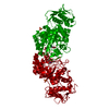
| ||||||||
| Unit cell |
| ||||||||
| Components on special symmetry positions |
|
-Components
-Protein / Sugars , 2 types, 2 molecules A
| #1: Protein | Hexosaminidase / beta-hexosaminidase / SpHEX Mass: 56125.793 Da / Num. of mol.: 1 / Mutation: D319N Source method: isolated from a genetically manipulated source Source: (gene. exp.) Streptomyces plicatus (bacteria) / Plasmid: pET3A / Species (production host): Escherichia coli / Production host: Escherichia coli BL21(DE3) (bacteria) / Strain (production host): BL21(DE3) / References: UniProt: O85361, beta-N-acetylhexosaminidase |
|---|---|
| #2: Sugar | ChemComp-NAG / N-Acetylglucosamine Type: D-saccharide, beta linking / Mass: 221.208 Da / Num. of mol.: 1 Type: D-saccharide, beta linking / Mass: 221.208 Da / Num. of mol.: 1Source method: isolated from a genetically manipulated source Formula: C8H15NO6 |
-Non-polymers , 4 types, 292 molecules 


| #3: Chemical | Chloride Mass: 35.453 Da / Num. of mol.: 3 / Source method: obtained synthetically / Formula: Cl Mass: 35.453 Da / Num. of mol.: 3 / Source method: obtained synthetically / Formula: Cl#4: Chemical | ChemComp-SO4 / | Sulfate Mass: 96.063 Da / Num. of mol.: 1 / Source method: obtained synthetically / Formula: SO4 Mass: 96.063 Da / Num. of mol.: 1 / Source method: obtained synthetically / Formula: SO4#5: Chemical | ChemComp-GOL / | Glycerol Mass: 92.094 Da / Num. of mol.: 1 / Source method: obtained synthetically / Formula: C3H8O3 Mass: 92.094 Da / Num. of mol.: 1 / Source method: obtained synthetically / Formula: C3H8O3#6: Water | ChemComp-HOH / | WaterMass: 18.015 Da / Num. of mol.: 287 / Source method: isolated from a natural source / Formula: H2O |
|---|
-Experimental details
-Experiment
| Experiment | Method: X-RAY DIFFRACTION / Number of used crystals: 1 |
|---|
- Sample preparation
| Crystal | Density Matthews: 4.05 Å3/Da / Density % sol: 69.6 % | |||||||||||||||||||||||||||||||||||||||||||||||||
|---|---|---|---|---|---|---|---|---|---|---|---|---|---|---|---|---|---|---|---|---|---|---|---|---|---|---|---|---|---|---|---|---|---|---|---|---|---|---|---|---|---|---|---|---|---|---|---|---|---|---|
| Crystal grow | Temperature: 298 K / Method: vapor diffusion, hanging drop / pH: 6 Details: AMMONIUM SULPHATE, TRI-SODIUM CITRATE, SODIUM CHLORIDE, GLYCEROL, NAG, pH 6.0, VAPOR DIFFUSION, HANGING DROP, temperature 298.0K | |||||||||||||||||||||||||||||||||||||||||||||||||
| Crystal grow | *PLUS | |||||||||||||||||||||||||||||||||||||||||||||||||
| Components of the solutions | *PLUS
|
-Data collection
| Diffraction | Mean temperature: 100 K |
|---|---|
| Diffraction source | Source: ROTATING ANODE / Type: RIGAKU / Wavelength: 1.54 Å |
| Detector | Type: RIGAKU RAXIS IV / Detector: IMAGE PLATE / Date: Aug 14, 2001 / Details: OSMIC optics |
| Radiation | Protocol: SINGLE WAVELENGTH / Monochromatic (M) / Laue (L): M / Scattering type: x-ray |
| Radiation wavelength | Wavelength: 1.54 Å / Relative weight: 1 |
| Reflection | Resolution: 1.95→40 Å / Num. all: 65848 / Num. obs: 65848 / % possible obs: 96.9 % / Observed criterion σ(I): -3 / Redundancy: 19 % / Biso Wilson estimate: 15.7 Å2 / Rsym value: 0.056 / Net I/σ(I): 30.5 |
| Reflection shell | Resolution: 1.95→2.02 Å / Redundancy: 19 % / Mean I/σ(I) obs: 5.5 / Rsym value: 0.256 / % possible all: 98.5 |
| Reflection | *PLUS Num. measured all: 1300022 / Rmerge(I) obs: 0.056 |
| Reflection shell | *PLUS % possible obs: 98.5 % / Num. unique obs: 6556 / Rmerge(I) obs: 0.256 |
- Processing
| Software |
| ||||||||||||||||||||||||||||||||||||||||||||||||||||||||||||||||||||||||||||||||
|---|---|---|---|---|---|---|---|---|---|---|---|---|---|---|---|---|---|---|---|---|---|---|---|---|---|---|---|---|---|---|---|---|---|---|---|---|---|---|---|---|---|---|---|---|---|---|---|---|---|---|---|---|---|---|---|---|---|---|---|---|---|---|---|---|---|---|---|---|---|---|---|---|---|---|---|---|---|---|---|---|---|
| Refinement | Method to determine structure: FOURIER SYNTHESIS Starting model: PDB ENTRY 1HP4 Resolution: 1.95→39.19 Å / Rfactor Rfree error: 0.003 / Isotropic thermal model: RESTRAINED / Cross valid method: THROUGHOUT / σ(F): 0 / Stereochemistry target values: Engh & Huber
| ||||||||||||||||||||||||||||||||||||||||||||||||||||||||||||||||||||||||||||||||
| Solvent computation | Solvent model: FLAT MODEL / Bsol: 43.3872 Å2 / ksol: 0.377555 e/Å3 | ||||||||||||||||||||||||||||||||||||||||||||||||||||||||||||||||||||||||||||||||
| Displacement parameters | Biso mean: 24.6 Å2
| ||||||||||||||||||||||||||||||||||||||||||||||||||||||||||||||||||||||||||||||||
| Refine analyze | Luzzati coordinate error free: 0.24 Å / Luzzati sigma a free: 0.16 Å | ||||||||||||||||||||||||||||||||||||||||||||||||||||||||||||||||||||||||||||||||
| Refinement step | Cycle: LAST / Resolution: 1.95→39.19 Å
| ||||||||||||||||||||||||||||||||||||||||||||||||||||||||||||||||||||||||||||||||
| Refine LS restraints |
| ||||||||||||||||||||||||||||||||||||||||||||||||||||||||||||||||||||||||||||||||
| LS refinement shell | Resolution: 1.95→2.02 Å / Rfactor Rfree error: 0.01 / Total num. of bins used: 6
| ||||||||||||||||||||||||||||||||||||||||||||||||||||||||||||||||||||||||||||||||
| Xplor file |
| ||||||||||||||||||||||||||||||||||||||||||||||||||||||||||||||||||||||||||||||||
| Refinement | *PLUS Lowest resolution: 39.2 Å / % reflection Rfree: 10 % | ||||||||||||||||||||||||||||||||||||||||||||||||||||||||||||||||||||||||||||||||
| Solvent computation | *PLUS | ||||||||||||||||||||||||||||||||||||||||||||||||||||||||||||||||||||||||||||||||
| Displacement parameters | *PLUS | ||||||||||||||||||||||||||||||||||||||||||||||||||||||||||||||||||||||||||||||||
| Refine LS restraints | *PLUS
|
