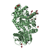
SHEET DETERMINATION METHOD: DSSP THE SHEETS PRESENTED AS "AC" IN EACH CHAIN ON SHEET RECORDS BELOW ... SHEET DETERMINATION METHOD: DSSP THE SHEETS PRESENTED AS "AC" IN EACH CHAIN ON SHEET RECORDS BELOW IS ACTUALLY AN 8-STRANDED BARREL THIS IS REPRESENTED BY A 9-STRANDED SHEET IN WHICH THE FIRST AND LAST STRANDS ARE IDENTICAL.
Mass: 18.015 Da / Num. of mol.: 454 / Source method: isolated from a natural source / Formula: H2O
Has protein modification
Y
Sequence details
THE FIRST 41 AMINO ACIDS ARE NOT PRESENT IN THE EXPRESSED, SECRETED PROTEIN, AND MAY CONTAIN A ...THE FIRST 41 AMINO ACIDS ARE NOT PRESENT IN THE EXPRESSED, SECRETED PROTEIN, AND MAY CONTAIN A POSSIBLE N-TERMINAL SIGNAL PEPTIDE SEQUENCE. RESIDUE 302 IS MUTATED TO GLN.
-
Experimental details
-
Experiment
Experiment
Method: X-RAY DIFFRACTION / Number of used crystals: 1
-
Sample preparation
Crystal
Density Matthews: 2.5 Å3/Da / Density % sol: 51 % / Description: NONE
Resolution: 1.8→71.83 Å / Cor.coef. Fo:Fc: 0.963 / Cor.coef. Fo:Fc free: 0.943 / SU B: 2.537 / SU ML: 0.078 / Cross valid method: THROUGHOUT / ESU R: 0.116 / ESU R Free: 0.116 / Stereochemistry target values: MAXIMUM LIKELIHOOD / Details: HYDROGENS HAVE BEEN ADDED IN THE RIDING POSITIONS.
Rfactor
Num. reflection
% reflection
Selection details
Rfree
0.21006
2622
5.2 %
RANDOM
Rwork
0.16709
-
-
-
obs
0.16926
48264
99.91 %
-
Solvent computation
Ion probe radii: 0.8 Å / Shrinkage radii: 0.8 Å / VDW probe radii: 1.2 Å / Solvent model: MASK
 Movie
Movie Controller

 Yorodumi
 Open data
 Basic information
 Components
 Keywords
 Function and homology information
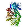
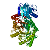
 STREPTOMYCES COELICOLOR (bacteria)
 X-RAY DIFFRACTION /
  Authors
 Citation
 Structure visualization
 Downloads & links
 Other downloads


 PDBj
PDBj
 Assembly
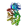
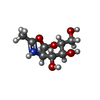
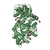
 Mass: 204.200 Da / Num. of mol.: 1 / Source method: obtained synthetically / Formula: C8H14NO5
 Mass: 62.068 Da / Num. of mol.: 11 / Source method: obtained synthetically / Formula: C2H6O2
 Mass: 18.015 Da / Num. of mol.: 454 / Source method: isolated from a natural source / Formula: H2O
 Sample preparation
 / Beamline: I04 / Wavelength: 0.9795
  Processing