 Movie
Movie Controller

[English] 日本語
 Yorodumi
- PDB-1lya: CRYSTAL STRUCTURES OF NATIVE AND INHIBITED FORMS OF HUMAN CATHEPS... -
+ Open data

- Basic information
| Entry | Database: PDB / ID: 1lya | |||||||||
|---|---|---|---|---|---|---|---|---|---|---|
| Title | CRYSTAL STRUCTURES OF NATIVE AND INHIBITED FORMS OF HUMAN CATHEPSIN D: IMPLICATIONS FOR LYSOSOMAL TARGETING AND DRUG DESIGN | |||||||||
 Components Components | (CATHEPSIN D Cathepsin) x 2 Cathepsin) x 2 | |||||||||
 Keywords Keywords | LYSOSOMAL ASPARTIC PROTEASE | |||||||||
| Function / homology |  Function and homology informationcathepsin D / aspartic-type peptidase activity / regulation of establishment of protein localization / lipoprotein catabolic process / insulin catabolic process / insulin receptor recycling / positive regulation of cysteine-type endopeptidase activity involved in apoptotic process / autophagosome assembly / Collagen degradation / Metabolism of Angiotensinogen to Angiotensins ...cathepsin D / aspartic-type peptidase activity / regulation of establishment of protein localization / lipoprotein catabolic process / insulin catabolic process / insulin receptor recycling / positive regulation of cysteine-type endopeptidase activity involved in apoptotic process / autophagosome assembly / Collagen degradation / Metabolism of Angiotensinogen to Angiotensins / Insulin receptor recycling / MHC class II antigen presentation / lysosomal lumen / endosome lumen / specific granule lumen / antigen processing and presentation of exogenous peptide antigen via MHC class II / melanosome / tertiary granule lumen / peptidase activity / collagen-containing extracellular matrix / Estrogen-dependent gene expression / ficolin-1-rich granule lumen / lysosome / aspartic-type endopeptidase activity / endosome membrane / positive regulation of apoptotic process / membrane raft / lysosomal membrane / cysteine-type endopeptidase activity / Neutrophil degranulation / proteolysis / extracellular space / extracellular exosome / extracellular region Function and homology informationcathepsin D / aspartic-type peptidase activity / regulation of establishment of protein localization / lipoprotein catabolic process / insulin catabolic process / insulin receptor recycling / positive regulation of cysteine-type endopeptidase activity involved in apoptotic process / autophagosome assembly / Collagen degradation / Metabolism of Angiotensinogen to Angiotensins ...cathepsin D / aspartic-type peptidase activity / regulation of establishment of protein localization / lipoprotein catabolic process / insulin catabolic process / insulin receptor recycling / positive regulation of cysteine-type endopeptidase activity involved in apoptotic process / autophagosome assembly / Collagen degradation / Metabolism of Angiotensinogen to Angiotensins / Insulin receptor recycling / MHC class II antigen presentation / lysosomal lumen / endosome lumen / specific granule lumen / antigen processing and presentation of exogenous peptide antigen via MHC class II / melanosome / tertiary granule lumen / peptidase activity / collagen-containing extracellular matrix / Estrogen-dependent gene expression / ficolin-1-rich granule lumen / lysosome / aspartic-type endopeptidase activity / endosome membrane / positive regulation of apoptotic process / membrane raft / lysosomal membrane / cysteine-type endopeptidase activity / Neutrophil degranulation / proteolysis / extracellular space / extracellular exosome / extracellular regionSimilarity search - Function | |||||||||
| Biological species |  Homo sapiens (human) Homo sapiens (human) | |||||||||
| Method | X-RAY DIFFRACTION / Resolution: 2.5 Å | |||||||||
 Authors Authors | Baldwin, E.T. / Bhat, T.N. / Gulnik, S. / Erickson, J.W. | |||||||||
 Citation Citation | Journal: Proc.Natl.Acad.Sci.USA / Year: 1993 Title: Crystal structures of native and inhibited forms of human cathepsin D: implications for lysosomal targeting and drug design. Authors: Baldwin, E.T. / Bhat, T.N. / Gulnik, S. / Hosur, M.V. / Sowder 2nd., R.C. / Cachau, R.E. / Collins, J. / Silva, A.M. / Erickson, J.W. #1: Journal: J.Mol.Biol. / Year: 1992Title: Human Liver Cathepsin D. Purification, Crystallization and Preliminary X-Ray Diffraction Analysis of a Lysosomal Enzyme Authors: Gulnik, S. / Baldwin, E.T. / Tarasova, N. / Erickson, J. | |||||||||
| History |
| |||||||||
| Remark 700 | SHEET THERE ARE SEVERAL BIFURCATED SHEETS IN THIS STRUCTURE. THESE ARE REPRESENTED BY TWO SHEETS ...SHEET THERE ARE SEVERAL BIFURCATED SHEETS IN THIS STRUCTURE. THESE ARE REPRESENTED BY TWO SHEETS WHICH HAVE ONE OR MORE IDENTICAL STRANDS. SHEETS *1A1* AND *1B1* AND SHEETS *1A2* AND *1B2* REPRESENT ONE BIFURCATED SHEET IN EACH MOLECULE. SHEETS *2A1* AND *2B1* AND *2A2* AND *2B2* REPRESENT ONE BIFURCATED SHEET IN EACH MOLECULE. |
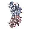
- Structure visualization
| Structure viewer | Molecule: MolmilJmol/JSmol |
|---|
- Downloads & links
-Download
| PDBx/mmCIF format | 1lya.cif.gz | 168.1 KB | Display | PDBx/mmCIF format |
|---|---|---|---|---|
| PDB format | pdb1lya.ent.gz | 137.8 KB | Display | PDB format |
| PDBx/mmJSON format | 1lya.json.gz | Tree view | PDBx/mmJSON format | |
| Others |  Other downloads Other downloads |
-Validation report
| Arichive directory | https://data.pdbj.org/pub/pdb/validation_reports/ly/1lyaftp://data.pdbj.org/pub/pdb/validation_reports/ly/1lya | HTTPS FTP |
|---|
-Related structure data


-Links
 PDBj
PDBj


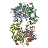
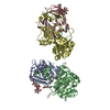
- Assembly
| Deposited unit | 
| ||||||||
|---|---|---|---|---|---|---|---|---|---|
| 1 |
| ||||||||
| 2 | 
| ||||||||
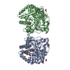
| Unit cell |
| ||||||||
| Atom site foot note | 1: CIS PROLINE - PRO A 24 / 2: CIS PROLINE - PRO A 95 / 3: CIS PROLINE - PRO B 177 / 4: CIS PROLINE - PRO B 314 / 5: CIS PROLINE - PRO B 317 / 6: CIS PROLINE - PRO C 2 / 7: CIS PROLINE - PRO C 24 / 8: CIS PROLINE - PRO C 95 / 9: CIS PROLINE - PRO D 177 10: PRO D 313 - PRO D 314 OMEGA = 36.80 PEPTIDE BOND DEVIATES SIGNIFICANTLY FROM TRANS CONFORMATION 11: CIS PROLINE - PRO D 317 | ||||||||
| Noncrystallographic symmetry (NCS) | NCS oper: (Code: given Matrix: (0.7353, 0.6747, -0.06415), Vector : Details | THE TRANSFORMATION PRESENTED ON *MTRIX* RECORDS BELOW WILL YIELD APPROXIMATE COORDINATES FOR CHAINS *A* AND *B* WHEN APPLIED TO CHAINS *C* AND *D*, RESPECTIVELY. | |
-Components
| #1: Protein | Cathepsin Mass: 10688.941 Da / Num. of mol.: 2 / Source method: isolated from a natural source / Source: (natural) Homo sapiens (human) / Organ: LIVER / Tissue: LIVER / References: UniProt: P07339, cathepsin D#2: Protein | CathepsinMass: 26270.207 Da / Num. of mol.: 2 / Source method: isolated from a natural source / Source: (natural) Homo sapiens (human) / Organ: LIVER / Tissue: LIVER / References: UniProt: P07339, cathepsin D#3: Polysaccharide | / Mass: 748.682 Da / Num. of mol.: 2Source method: isolated from a genetically manipulated source #4: Sugar | N-Acetylglucosamine  Type: D-saccharide, beta linking / Mass: 221.208 Da / Num. of mol.: 2 Type: D-saccharide, beta linking / Mass: 221.208 Da / Num. of mol.: 2Source method: isolated from a genetically manipulated source Formula: C8H15NO6 #5: Water | ChemComp-HOH / | Water Mass: 18.015 Da / Num. of mol.: 46 / Source method: isolated from a natural source / Formula: H2O Mass: 18.015 Da / Num. of mol.: 46 / Source method: isolated from a natural source / Formula: H2O |
|---|
-Experimental details
-Experiment
| Experiment | Method: X-RAY DIFFRACTION |
|---|
- Sample preparation
| Crystal | Density Matthews: 3.22 Å3/Da / Density % sol: 61.8 % | ||||||||||||||||||||
|---|---|---|---|---|---|---|---|---|---|---|---|---|---|---|---|---|---|---|---|---|---|
| Crystal grow | *PLUS Temperature: 20 ℃ / pH: 5.1 / Method: vapor diffusion, hanging dropDetails: taken from Gulnik, S. et al (1992). J. Mol. Biol., 227, 265-270. | ||||||||||||||||||||
| Components of the solutions | *PLUS
|
-Data collection
| Radiation | Scattering type: x-ray |
|---|---|
| Radiation wavelength | Relative weight: 1 |
- Processing
| Software |
| ||||||||||||||||||||||||||||||||||||||||||||||||||||||||||||
|---|---|---|---|---|---|---|---|---|---|---|---|---|---|---|---|---|---|---|---|---|---|---|---|---|---|---|---|---|---|---|---|---|---|---|---|---|---|---|---|---|---|---|---|---|---|---|---|---|---|---|---|---|---|---|---|---|---|---|---|---|---|
| Refinement | Resolution: 2.5→10 Å Details: THERE ARE TWO N-LINKED OLIGOSACCHARIDES ATTACHED AT RESIDUES ASN 70 AND ASN 199 OF EACH MOLECULE. FOUR SUGARS WERE INCLUDED FOR REFINEMENT AT ASN 70 AND A SINGLE N-LINKED NAG AT ASN 199. THE ...Details: THERE ARE TWO N-LINKED OLIGOSACCHARIDES ATTACHED AT RESIDUES ASN 70 AND ASN 199 OF EACH MOLECULE. FOUR SUGARS WERE INCLUDED FOR REFINEMENT AT ASN 70 AND A SINGLE N-LINKED NAG AT ASN 199. THE SUGARS ARE LINKED AS FOLLOWS: ASN A 70 -- NAG A 1 -- NAG A 2 -- MAN A 3 -- MAN A 8 ASN B 199 -- NAG B 1 ASN C 70 -- NAG C 1 -- NAG C 2 -- MAN C 3 -- MAN C 8 ASN D 199 -- NAG D 1
| ||||||||||||||||||||||||||||||||||||||||||||||||||||||||||||
| Refinement step | Cycle: LAST / Resolution: 2.5→10 Å
| ||||||||||||||||||||||||||||||||||||||||||||||||||||||||||||
| Refine LS restraints |
| ||||||||||||||||||||||||||||||||||||||||||||||||||||||||||||
| Software | *PLUS Name: X-PLOR / Classification: refinement | ||||||||||||||||||||||||||||||||||||||||||||||||||||||||||||
| Refinement | *PLUS Highest resolution: 2.5 Å / Lowest resolution: 10 Å / Num. reflection obs: 28077 / Rfactor obs: 0.188 | ||||||||||||||||||||||||||||||||||||||||||||||||||||||||||||
| Solvent computation | *PLUS | ||||||||||||||||||||||||||||||||||||||||||||||||||||||||||||
| Displacement parameters | *PLUS | ||||||||||||||||||||||||||||||||||||||||||||||||||||||||||||
| Refine LS restraints | *PLUS
|
