 Movie
Movie Controller

[English] 日本語
 Yorodumi
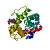
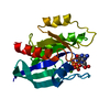
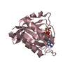
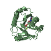
- PDB-1lsp: THE CRYSTAL STRUCTURE OF A BULGECIN-INHIBITED G-TYPE LYSOZYME FRO... -
+ Open data

- Basic information
| Entry | Database: PDB / ID: 1lsp | ||||||
|---|---|---|---|---|---|---|---|
| Title | THE CRYSTAL STRUCTURE OF A BULGECIN-INHIBITED G-TYPE LYSOZYME FROM THE EGG-WHITE OF THE AUSTRALIAN BLACK SWAN. A COMPARISON OF THE BINDING OF BULGECIN TO THREE MURAMIDASES | ||||||
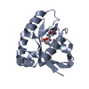
 Components Components | LYSOZYME | ||||||
 Keywords Keywords | HYDROLASE (O-GLYCOSYL) | ||||||
| Function / homology |  Function and homology information Function and homology informationpeptidoglycan catabolic process / lysozyme / lysozyme activity / killing of cells of another organism / defense response to Gram-positive bacterium / extracellular region Similarity search - Function | ||||||
| Biological species |  Cygnus atratus (black swan) Cygnus atratus (black swan) | ||||||
| Method |  X-RAY DIFFRACTION / Resolution: 2.45 Å X-RAY DIFFRACTION / Resolution: 2.45 Å | ||||||
 Authors Authors | Karlsen, S. / Rao, Z.H. / Hough, E. / Isaacs, N.W. | ||||||
 Citation Citation | Journal: Acta Crystallogr.,Sect.D / Year: 1996 Title: Structure of a bulgecin-inhibited g-type lysozyme from the egg white of the Australian black swan. A comparison of the binding of bulgecin to three muramidases. Authors: Karlsen, S. / Hough, E. / Rao, Z.H. / Isaacs, N.W. #1: Journal: To be PublishedTitle: The Crystal Structure of a Complex between Bulgecin, a Bacterial Metabolite, and Lysozyme from the Rainbow Trout Authors: Karlsen, S. / Hough, E. #2: Journal: Aust.J.Biol.Sci. / Year: 1985Title: Three-Dimensional Structure of Goose-Type Lysozyme from the Egg White of the Australian Black Swan, Cygnus Atratus Authors: Isaacs, N.W. / Machin, K.J. / Masakuni, M. #3: Journal: Tetrahedron / Year: 1984Title: Structures of Bulgecins, Bacterial Metabolites with Bulge-Inducing Activity Authors: Shinagawa, S. / Kasahara, F. / Wada, Y. / Harada, S. / Asai, M. | ||||||
| History |
|
- Structure visualization
| Structure viewer | Molecule: MolmilJmol/JSmol |
|---|
- Downloads & links
-Download
| PDBx/mmCIF format | 1lsp.cif.gz | 52.1 KB | Display | PDBx/mmCIF format |
|---|---|---|---|---|
| PDB format | pdb1lsp.ent.gz | 36.4 KB | Display | PDB format |
| PDBx/mmJSON format | 1lsp.json.gz | Tree view | PDBx/mmJSON format | |
| Others |  Other downloads Other downloads |
-Validation report
| Arichive directory | https://data.pdbj.org/pub/pdb/validation_reports/ls/1lspftp://data.pdbj.org/pub/pdb/validation_reports/ls/1lsp | HTTPS FTP |
|---|
-Related structure data
| Similar structure data |
|---|

-Links
 PDBj
PDBj- Assembly
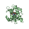
| Deposited unit | 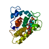
| ||||||||
|---|---|---|---|---|---|---|---|---|---|
| 1 |
| ||||||||
| Unit cell |
|
-Components
| #1: Protein | Mass: 20434.172 Da / Num. of mol.: 1 / Source method: isolated from a natural source / Source: (natural) Cygnus atratus (black swan) / Cellular location: EGG WHITE / References: UniProt: P00717, lysozyme |
|---|---|
| #2: Chemical | ChemComp-BUL / 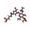  Mass: 551.543 Da / Num. of mol.: 1 / Source method: obtained synthetically / Formula: C16H29N3O14S2 Mass: 551.543 Da / Num. of mol.: 1 / Source method: obtained synthetically / Formula: C16H29N3O14S2 |
| #3: Water | ChemComp-HOH /  Mass: 18.015 Da / Num. of mol.: 109 / Source method: isolated from a natural source / Formula: H2O Mass: 18.015 Da / Num. of mol.: 109 / Source method: isolated from a natural source / Formula: H2O |
| Has protein modification | Y |
-Experimental details
-Experiment
| Experiment | Method: X-RAY DIFFRACTION / Number of used crystals: 1 |
|---|
- Sample preparation
| Crystal | Density Matthews: 2.8 Å3/Da / Density % sol: 55.99 % | ||||||||||||||||||
|---|---|---|---|---|---|---|---|---|---|---|---|---|---|---|---|---|---|---|---|
| Crystal grow | *PLUS pH: 6.5 / Method: vapor diffusion, hanging drop | ||||||||||||||||||
| Components of the solutions | *PLUS
|
-Data collection
| Diffraction source | Wavelength: 1.5418 Å |
|---|---|
| Detector | Type: SIEMENS / Detector: AREA DETECTOR / Date: Jun 1, 1994 |
| Radiation | Monochromatic (M) / Laue (L): M / Scattering type: x-ray |
| Radiation wavelength | Wavelength: 1.5418 Å / Relative weight: 1 |
| Reflection | Num. obs: 6959 / % possible obs: 78.9 % / Observed criterion σ(I): 3 / Rmerge(I) obs: 0.055 |
| Reflection | *PLUS Highest resolution: 2.45 Å / Lowest resolution: 32.94 Å |
- Processing
| Software |
| ||||||||||||||||||||||||||||||||||||||||||||||||||||||||||||||||||||||||||||||||||||
|---|---|---|---|---|---|---|---|---|---|---|---|---|---|---|---|---|---|---|---|---|---|---|---|---|---|---|---|---|---|---|---|---|---|---|---|---|---|---|---|---|---|---|---|---|---|---|---|---|---|---|---|---|---|---|---|---|---|---|---|---|---|---|---|---|---|---|---|---|---|---|---|---|---|---|---|---|---|---|---|---|---|---|---|---|---|
| Refinement | Resolution: 2.45→8 Å / σ(F): 3 /
| ||||||||||||||||||||||||||||||||||||||||||||||||||||||||||||||||||||||||||||||||||||
| Refine analyze | Luzzati coordinate error obs: 0.17 Å | ||||||||||||||||||||||||||||||||||||||||||||||||||||||||||||||||||||||||||||||||||||
| Refinement step | Cycle: LAST / Resolution: 2.45→8 Å
| ||||||||||||||||||||||||||||||||||||||||||||||||||||||||||||||||||||||||||||||||||||
| Refine LS restraints |
|
