 Movie
Movie Controller

[English] 日本語
 Yorodumi
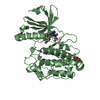
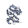
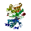
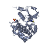
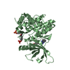
- PDB-1lf2: CRYSTAL STRUCTURE OF PLASMEPSIN II FROM P FALCIPARUM IN COMPLEX W... -
+ Open data

- Basic information
| Entry | Database: PDB / ID: 1lf2 | ||||||
|---|---|---|---|---|---|---|---|
| Title | CRYSTAL STRUCTURE OF PLASMEPSIN II FROM P FALCIPARUM IN COMPLEX WITH INHIBITOR RS370 | ||||||
 Components Components | Plasmepsin 2 | ||||||
 Keywords Keywords | HYDROLASE / plasmepsin / plasmodium falciparum / aspartic protease | ||||||
| Function / homology |  Function and homology information Function and homology informationcytostome / plasmepsin II / acquisition of nutrients from host / vacuolar lumen / food vacuole / vacuolar membrane / aspartic-type endopeptidase activity / proteolysis Similarity search - Function | ||||||
| Biological species |  | ||||||
| Method |  X-RAY DIFFRACTION / SYNCHROTRON / MOLECULAR REPLACEMENT / Resolution: 1.8 Å X-RAY DIFFRACTION / SYNCHROTRON / MOLECULAR REPLACEMENT / Resolution: 1.8 Å | ||||||
 Authors Authors | Asojo, O.A. / Afonina, E. / Gulnik, S.V. / Yu, B. / Erickson, J.W. / Randad, R. / Mehadjed, D. / Silva, A.M. | ||||||
 Citation Citation | Journal: Acta Crystallogr.,Sect.D / Year: 2002 Title: Structures of Ser205 mutant plasmepsin II from Plasmodium falciparum at 1.8 A in complex with the inhibitors rs367 and rs370. Authors: Asojo, O.A. / Afonina, E. / Gulnik, S.V. / Yu, B. / Erickson, J.W. / Randad, R. / Medjahed, D. / Silva, A.M. #1: Journal: J.Mol.Biol. / Year: 2003Title: Novel uncomplexed and complexed structures of plasmepsin II, an aspartic protease from Plasmodium falciparum. Authors: Asojo, O.A. / Gulnik, S.V. / Afonina, E. / Yu, B. / Ellman, J.A. / Haque, T.S. / Silva, A.M. | ||||||
| History |
|
- Structure visualization
| Structure viewer | Molecule: MolmilJmol/JSmol |
|---|
- Downloads & links
-Download
| PDBx/mmCIF format | 1lf2.cif.gz | 87 KB | Display | PDBx/mmCIF format |
|---|---|---|---|---|
| PDB format | pdb1lf2.ent.gz | 65 KB | Display | PDB format |
| PDBx/mmJSON format | 1lf2.json.gz | Tree view | PDBx/mmJSON format | |
| Others |  Other downloads Other downloads |
-Validation report
| Arichive directory | https://data.pdbj.org/pub/pdb/validation_reports/lf/1lf2ftp://data.pdbj.org/pub/pdb/validation_reports/lf/1lf2 | HTTPS FTP |
|---|
-Related structure data


-Links
 PDBj
PDBj- Assembly
| Deposited unit | 
| ||||||||
|---|---|---|---|---|---|---|---|---|---|
| 1 |
| ||||||||
| Unit cell |
|
-Components
| #1: Protein | Mass: 37079.824 Da / Num. of mol.: 1 / Fragment: Residues 123-453 Source method: isolated from a genetically manipulated source Source: (gene. exp.) Plasmid: PET 22B (NOVAGEN) / Production host:  |
|---|---|
| #2: Chemical | ChemComp-R37 / 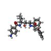  Mass: 531.686 Da / Num. of mol.: 1 / Source method: obtained synthetically / Formula: C32H41N3O4 Mass: 531.686 Da / Num. of mol.: 1 / Source method: obtained synthetically / Formula: C32H41N3O4 |
| #3: Water | ChemComp-HOH /  Mass: 18.015 Da / Num. of mol.: 338 / Source method: isolated from a natural source / Formula: H2O Mass: 18.015 Da / Num. of mol.: 338 / Source method: isolated from a natural source / Formula: H2O |
| Has protein modification | Y |
-Experimental details
-Experiment
| Experiment | Method: X-RAY DIFFRACTION / Number of used crystals: 1 |
|---|
- Sample preparation
| Crystal | Density Matthews: 2.67 Å3/Da / Density % sol: 53.95 % | ||||||||||||||||||||||||||||||||||||||||||||||||||||||
|---|---|---|---|---|---|---|---|---|---|---|---|---|---|---|---|---|---|---|---|---|---|---|---|---|---|---|---|---|---|---|---|---|---|---|---|---|---|---|---|---|---|---|---|---|---|---|---|---|---|---|---|---|---|---|---|
| Crystal grow | *PLUS Temperature: 293 K / pH: 7 / Method: vapor diffusion, hanging drop | ||||||||||||||||||||||||||||||||||||||||||||||||||||||
| Components of the solutions | *PLUS
|
-Data collection
| Diffraction | Mean temperature: 90 K |
|---|---|
| Diffraction source | Source: SYNCHROTRON / Site: NSLS  / Beamline: X9B / Wavelength: 0.92 / Beamline: X9B / Wavelength: 0.92 |
| Detector | Type: MAR scanner 345 mm plate / Detector: IMAGE PLATE / Date: Nov 15, 1998 |
| Radiation | Protocol: SINGLE WAVELENGTH / Monochromatic (M) / Laue (L): M / Scattering type: x-ray |
| Radiation wavelength | Wavelength: 0.92 Å / Relative weight: 1 |
| Reflection | Resolution: 1.8→20 Å / Num. obs: 35627 / % possible obs: 95 % / Observed criterion σ(I): 2 / Redundancy: 4 % / Rmerge(I) obs: 0.058 / Net I/σ(I): 13.1 |
| Reflection shell | Resolution: 1.8→1.9 Å / Redundancy: 3 % / Rmerge(I) obs: 0.25 / Mean I/σ(I) obs: 2 / % possible all: 53 |
| Reflection | *PLUS Lowest resolution: 20 Å / % possible obs: 95 % |
| Reflection shell | *PLUS % possible obs: 53 % / Rmerge(I) obs: 0.25 / Mean I/σ(I) obs: 3 |
- Processing
| Software |
| |||||||||||||||||||||
|---|---|---|---|---|---|---|---|---|---|---|---|---|---|---|---|---|---|---|---|---|---|---|
| Refinement | Method to determine structure: MOLECULAR REPLACEMENT / Resolution: 1.8→20 Å / σ(F): 2
| |||||||||||||||||||||
| Displacement parameters | Biso mean: 35.4 Å2 | |||||||||||||||||||||
| Refinement step | Cycle: LAST / Resolution: 1.8→20 Å
| |||||||||||||||||||||
| Refine LS restraints |
| |||||||||||||||||||||
| Refinement | *PLUS Lowest resolution: 20 Å / % reflection Rfree: 5 % | |||||||||||||||||||||
| Solvent computation | *PLUS | |||||||||||||||||||||
| Displacement parameters | *PLUS | |||||||||||||||||||||
| Refine LS restraints | *PLUS
| |||||||||||||||||||||
| LS refinement shell | *PLUS Highest resolution: 1.8 Å / Lowest resolution: 1.9 Å / Rfactor Rfree: 0.287 / Rfactor Rwork: 0.267 |
