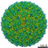
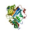
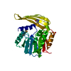
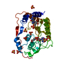
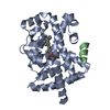
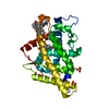
[English] 日本語
Yorodumi
- PDB-1l9k: dengue methyltransferase -

+
Open data


ID or keywords:

Loading...

-
Basic information

Entry
Database: PDB / ID: 1l9k
Titledengue methyltransferase
ComponentsRNA-DIRECTED RNA POLYMERASERNA-dependent RNA polymerase
KeywordsTRANSFERASE / methyltransferase fold / complex with s-adenosyl-l-homocysteine
Function / homology
Function and homology information


symbiont-mediated suppression of host JAK-STAT cascade via inhibition of host TYK2 activity / flavivirin / host cell mitochondrion / symbiont-mediated suppression of host JAK-STAT cascade via inhibition of STAT2 activity / symbiont-mediated suppression of host cytoplasmic pattern recognition receptor signaling pathway via inhibition of MAVS activity / viral capsid / : / nucleoside-triphosphate phosphatase / double-stranded RNA binding / protein complex oligomerization ...symbiont-mediated suppression of host JAK-STAT cascade via inhibition of host TYK2 activity / flavivirin / host cell mitochondrion / symbiont-mediated suppression of host JAK-STAT cascade via inhibition of STAT2 activity / symbiont-mediated suppression of host cytoplasmic pattern recognition receptor signaling pathway via inhibition of MAVS activity / viral capsid / : / nucleoside-triphosphate phosphatase / double-stranded RNA binding / protein complex oligomerization / monoatomic ion channel activity / mRNA (guanine-N7)-methyltransferase / methyltransferase cap1 / clathrin-dependent endocytosis of virus by host cell / mRNA (nucleoside-2'-O-)-methyltransferase activity / mRNA 5'-cap (guanine-N7-)-methyltransferase activity / RNA helicase activity / host cell endoplasmic reticulum membrane / host cell perinuclear region of cytoplasm / protein dimerization activity / RNA helicase / induction by virus of host autophagy / RNA-directed RNA polymerase / viral RNA genome replication / RNA-dependent RNA polymerase activity / serine-type endopeptidase activity / fusion of virus membrane with host endosome membrane / viral envelope / symbiont-mediated suppression of host type I interferon-mediated signaling pathway / host cell nucleus / virion attachment to host cell / virion membrane / structural molecule activity / ATP hydrolysis activity / proteolysis / extracellular region / ATP binding / membrane / metal ion binding
Similarity search - Function
: / Flavivirus envelope glycoprotein E, stem/anchor domain / RNA-directed RNA polymerase, thumb domain, Flavivirus / Flavivirus RNA-directed RNA polymerase, thumb domain / Flavivirus capsid protein C superfamily / : / Flavivirus non-structural protein NS2B / Flavivirus NS3 helicase, C-terminal helical domain / Genome polyprotein, Flavivirus / Flavivirus non-structural protein NS4A ...: / Flavivirus envelope glycoprotein E, stem/anchor domain / RNA-directed RNA polymerase, thumb domain, Flavivirus / Flavivirus RNA-directed RNA polymerase, thumb domain / Flavivirus capsid protein C superfamily / : / Flavivirus non-structural protein NS2B / Flavivirus NS3 helicase, C-terminal helical domain / Genome polyprotein, Flavivirus / Flavivirus non-structural protein NS4A / Flavivirus non-structural protein NS2B / Flavivirus capsid protein C / Flavivirus non-structural protein NS4B / mRNA cap 0/1 methyltransferase / Flavivirus capsid protein C / Flavivirus non-structural protein NS4B / Flavivirus non-structural protein NS4A / Flavivirus NS2B domain profile. / mRNA cap 0 and cap 1 methyltransferase (EC 2.1.1.56 and EC 2.1.1.57) domain profile. / Flavivirus non-structural protein NS2A / Flavivirus non-structural protein NS2A / Flavivirus NS3, petidase S7 / Peptidase S7, Flavivirus NS3 serine protease / Flavivirus NS3 protease (NS3pro) domain profile. / Envelope glycoprotein M, flavivirus / Flavivirus envelope glycoprotein M / RNA-directed RNA polymerase, flavivirus / Flavivirus RNA-directed RNA polymerase, fingers and palm domains / Flavivirus non-structural Protein NS1 / Flavivirus non-structural protein NS1 / Envelope glycoprotein M superfamily, flavivirus / Flavivirus polyprotein propeptide / Flavivirus polyprotein propeptide superfamily / Flavivirus polyprotein propeptide / Flaviviral glycoprotein E, central domain, subdomain 1 / Flaviviral glycoprotein E, central domain, subdomain 2 / Flavivirus envelope glycoprotein E, Stem/Anchor domain / Flavivirus glycoprotein E, immunoglobulin-like domain / Flavivirus envelope glycoprotein E, Stem/Anchor domain superfamily / Flavivirus glycoprotein, immunoglobulin-like domain / Flavivirus glycoprotein central and dimerisation domain / Flavivirus glycoprotein, central and dimerisation domains / Ribosomal RNA methyltransferase, FtsJ domain / FtsJ-like methyltransferase / Flavivirus/Alphavirus glycoprotein, immunoglobulin-like domain superfamily / Flavivirus glycoprotein, central and dimerisation domain superfamily / Flaviviral glycoprotein E, dimerisation domain / DEAD box, Flavivirus / Flavivirus DEAD domain / Vaccinia Virus protein VP39 / helicase superfamily c-terminal domain / Immunoglobulin E-set / Superfamilies 1 and 2 helicase C-terminal domain profile. / Superfamilies 1 and 2 helicase ATP-binding type-1 domain profile. / DEAD-like helicases superfamily / Helicase, C-terminal / Helicase superfamily 1/2, ATP-binding domain / RNA-directed RNA polymerase, catalytic domain / RdRp of positive ssRNA viruses catalytic domain profile. / S-adenosyl-L-methionine-dependent methyltransferase superfamily / Peptidase S1, PA clan, chymotrypsin-like fold / Peptidase S1, PA clan / DNA/RNA polymerase superfamily / P-loop containing nucleoside triphosphate hydrolase / Rossmann fold / 3-Layer(aba) Sandwich / Alpha Beta
Similarity search - Domain/homology
S-ADENOSYL-L-HOMOCYSTEINE / Genome polyprotein
Similarity search - Component
Biological speciesDengue virus
MethodX-RAY DIFFRACTION / SYNCHROTRON / MAD / Resolution: 2.4 Å
AuthorsEgloff, M.P. / Benarroch, D. / Selisko, B. / Romette, J.L. / Canard, B.
CitationJournal: Embo J. / Year: 2002
Title: An RNA cap (nucleoside-2'-O-) methyltransferase in the flavivirus RNA polymerase NS5: crystal structure and functional characterization
Authors: Egloff, M.P. / Benarroch, D. / Selisko, B. / Romette, J.L. / Canard, B.
History
DepositionMar 25, 2002Deposition site: RCSB / Processing site: RCSB
Revision 1.0Mar 25, 2003Provider: repository / Type: Initial release
Revision 1.1Apr 28, 2008Group: Version format compliance
Revision 1.2Jul 13, 2011Group: Source and taxonomy / Version format compliance
Revision 1.3Oct 27, 2021Group: Database references / Derived calculations / Category: database_2 / struct_ref_seq_dif / struct_site
Item: _database_2.pdbx_DOI / _database_2.pdbx_database_accession ..._database_2.pdbx_DOI / _database_2.pdbx_database_accession / _struct_ref_seq_dif.details / _struct_site.pdbx_auth_asym_id / _struct_site.pdbx_auth_comp_id / _struct_site.pdbx_auth_seq_id
Revision 1.4Feb 14, 2024Group: Data collection / Category: chem_comp_atom / chem_comp_bond

-
Structure visualization

Structure viewerMolecule:
MolmilJmol/JSmol

Downloads & links

-
Assembly

Deposited unit
A: RNA-DIRECTED RNA POLYMERASE
hetero molecules


Theoretical massNumber of molelcules
Total (without water)35,2627
Polymers34,3971
Non-polymers8656
Water1,17165
1


  • Idetical with deposited unit
  • defined by author&software
TypeNameSymmetry operationNumber
identity operation1_555x,y,z1
MethodPISA
2
A: RNA-DIRECTED RNA POLYMERASE
hetero molecules

A: RNA-DIRECTED RNA POLYMERASE
hetero molecules


Theoretical massNumber of molelcules
Total (without water)70,52414
Polymers68,7942
Non-polymers1,72912
Water362
TypeNameSymmetry operationNumber
identity operation1_555x,y,z1
crystal symmetry operation6_555-x,-x+y,-z+1/31
Buried area4930 Å2
ΔGint-161 kcal/mol
Surface area23710 Å2
MethodPISA
Unit cell
Length a, b, c (Å)111.560, 111.560, 56.330
Angle α, β, γ (deg.)90.00, 90.00, 120.00
Int Tables number152
Space group name H-MP3121

-
Components

#1: Protein RNA-DIRECTED RNA POLYMERASE / RNA-dependent RNA polymerase / methyltransferase


Mass: 34397.027 Da / Num. of mol.: 1 / Fragment: NS5 N-terminal domain / Mutation: V135I, K139R, N173S, P188S, R193K, T196A, Y201H
Source method: isolated from a genetically manipulated source
Source: (gene. exp.) Dengue virus / Genus: Flavivirus / Strain: 2 / Gene: NS5 New Guinea C / Plasmid: pQE30 / Production host: Escherichia coli (E. coli) / Strain (production host): BL21[pREP4] / References: UniProt: P12823, RNA-directed RNA polymerase
#2: Chemical
ChemComp-SO4 / SULFATE ION / Sulfate


Mass: 96.063 Da / Num. of mol.: 5 / Source method: obtained synthetically / Formula: SO4
#3: Chemical ChemComp-SAH / S-ADENOSYL-L-HOMOCYSTEINE / S-Adenosyl-L-homocysteine


Type: L-peptide linking / Mass: 384.411 Da / Num. of mol.: 1 / Source method: obtained synthetically / Formula: C14H20N6O5S
#4: Water ChemComp-HOH / water / Water


Mass: 18.015 Da / Num. of mol.: 65 / Source method: isolated from a natural source / Formula: H2O

-
Experimental details

-
Experiment

ExperimentMethod: X-RAY DIFFRACTION / Number of used crystals: 1

-
Sample preparation

CrystalDensity Matthews: 2.94 Å3/Da / Density % sol: 63 %
Crystal growTemperature: 291 K / Method: vapor diffusion, hanging drop / pH: 5.8
Details: sodium citrate 100mM, lithium sulfate 1.2 M, ammonium sulfate 0.5 M, pH 5.8, VAPOR DIFFUSION, HANGING DROP, temperature 291K
Crystal grow
*PLUS
Components of the solutions
*PLUS
IDConc.Common nameCrystal-IDSol-IDDetails
122 mg/mlprotein1drop
20.1 Msodium citrate1reservoirpH5.8
31.2 Mlithium sulfate1reservoir
40.5 Mammonium sulfate1reservoir

-
Data collection

Diffraction
IDMean temperature (K)Crystal-ID
11001
21001
1,21
Diffraction source
SourceSiteBeamlineIDWavelength (Å)
SYNCHROTRONESRF BM1410.83211, 1.00474, 1.00850
SYNCHROTRONESRF ID14-220.933
Detector
TypeIDDetectorDateDetails
MARRESEARCH1CCDSep 9, 2000mirror
ADSC QUANTUM 42CCDSep 10, 2000mirror
Radiation
IDMonochromatorProtocolMonochromatic (M) / Laue (L)Scattering typeWavelength-ID
1Si [111] channelMADMx-ray1
2diamond [111] crystalSINGLE WAVELENGTHMx-ray1
Radiation wavelength
IDWavelength (Å)Relative weight
10.832111
21.004741
31.00851
40.9331
ReflectionResolution: 2.4→27.96 Å / Num. all: 16248 / Num. obs: 16035 / % possible obs: 99.8 % / Observed criterion σ(F): 1 / Observed criterion σ(I): 0 / Redundancy: 5.5 % / Biso Wilson estimate: 50.8 Å2 / Limit h max: 40 / Limit h min: 0 / Limit k max: 40 / Limit k min: 0 / Limit l max: 23 / Limit l min: 0 / Observed criterion F max: 924791.66 / Observed criterion F min: 9 / Rmerge(I) obs: 0.062 / Rsym value: 0.051 / Net I/σ(I): 17.4
Reflection shellResolution: 2.4→2.53 Å / Redundancy: 5.2 % / Rmerge(I) obs: 0.432 / Mean I/σ(I) obs: 1.7 / Num. unique all: 2178 / Rsym value: 0.358 / % possible all: 99.6
Reflection
*PLUS
Highest resolution: 2.4 Å / Lowest resolution: 30 Å / Num. obs: 16248 / % possible obs: 99.6 % / Rmerge(I) obs: 0.051
Reflection shell
*PLUS
% possible obs: 99.6 % / Rmerge(I) obs: 0.358

-
Processing

Software
NameVersionClassificationNB
CNS1refinement
DENZOdata reduction
CCP4(SCALA)data scaling
MLPHAREphasing
RefinementMethod to determine structure: MAD / Resolution: 2.4→27.96 Å / Rfactor Rfree error: 0.008 / Occupancy max: 1 / Occupancy min: 1 / Cross valid method: THROUGHOUT / σ(F): 1 / Stereochemistry target values: Engh & Huber
RfactorNum. reflection% reflectionSelection details
Rfree0.257 945 5.9 %random
Rwork0.231 ---
all0.275 16049 --
obs0.275 16035 99.9 %-
Solvent computationSolvent model: CNS bulk solvent model used / Bsol: 36.9147 Å2 / ksol: 0.366544 e/Å3
Displacement parametersBiso max: 103.94 Å2 / Biso mean: 49.2 Å2 / Biso min: 29.86 Å2
Baniso -1Baniso -2Baniso -3
1-5.92 Å24.39 Å20 Å2
2--5.92 Å20 Å2
3----11.84 Å2
Refine analyze
FreeObs
Luzzati coordinate error0.35 Å0.32 Å
Luzzati d res low-5 Å
Luzzati sigma a0.37 Å0.37 Å
Luzzati d res high-2.4
Refinement stepCycle: LAST / Resolution: 2.4→27.96 Å
ProteinNucleic acidLigandSolventTotal
Num. atoms2042 0 51 65 2158
Refine LS restraints
Refine-IDTypeDev ideal
X-RAY DIFFRACTIONx_bond_d0.01
X-RAY DIFFRACTIONx_angle_deg1.4
X-RAY DIFFRACTIONx_torsion_deg23.8
X-RAY DIFFRACTIONx_torsion_impr_deg0.97
LS refinement shell

Refine-ID: X-RAY DIFFRACTION / Total num. of bins used: 8

Resolution (Å)Rfactor RfreeNum. reflection Rfree% reflection Rfree (%)Rfactor RworkNum. reflection RworkRfactor Rfree errorNum. reflection allNum. reflection obs% reflection obs (%)
2.4-2.510.3071005.10.30718600.0311961196099.9
2.51-2.640.3071135.70.30618650.0291985197899.6
2.64-2.810.2831175.90.28218800.0261998199799.9
2.81-3.020.2511135.70.25118650.0241984197899.7
3.02-3.330.25212060.25218810.0232006200199.8
3.33-3.810.2171195.90.21718840.0220042003100
3.81-4.790.1931306.40.19418960.01720262026100
4.79-27.960.2211336.30.2219590.0192097209299.8
Xplor file
Refine-IDSerial noParam fileTopol file
X-RAY DIFFRACTION1protein_rep.paramprotein.top
X-RAY DIFFRACTION2dna-rna_rep.paramdna-rna.top
X-RAY DIFFRACTION3water_rep.paramwater.top
X-RAY DIFFRACTION4ion.paramion.top
X-RAY DIFFRACTION5sah.paramsah.top
Software
*PLUS
Name: CNS / Classification: refinement
Refinement
*PLUS
Highest resolution: 2.4 Å / Lowest resolution: 30 Å / % reflection Rfree: 5 % / Rfactor Rfree: 0.25 / Rfactor Rwork: 0.236
Solvent computation
*PLUS
Displacement parameters
*PLUS
Refine LS restraints
*PLUS
Refine-IDTypeDev ideal
X-RAY DIFFRACTIONc_bond_d0.009
X-RAY DIFFRACTIONc_angle_deg1.485

+
About Yorodumi

-
News

-
Feb 9, 2022. New format data for meta-information of EMDB entries

New format data for meta-information of EMDB entries

  • Version 3 of the EMDB header file is now the official format.
  • The previous official version 1.9 will be removed from the archive.

Related info.:EMDB header

External links:wwPDB to switch to version 3 of the EMDB data model

-
Aug 12, 2020. Covid-19 info

Covid-19 info

URL: https://pdbj.org/emnavi/covid19.php

New page: Covid-19 featured information page in EM Navigator.

Related info.:Covid-19 info / Mar 5, 2020. Novel coronavirus structure data

+
Mar 5, 2020. Novel coronavirus structure data

Novel coronavirus structure data

Related info.:Yorodumi Speices / Aug 12, 2020. Covid-19 info

External links:COVID-19 featured content - PDBj / Molecule of the Month (242):Coronavirus Proteases

+
Jan 31, 2019. EMDB accession codes are about to change! (news from PDBe EMDB page)

EMDB accession codes are about to change! (news from PDBe EMDB page)

  • The allocation of 4 digits for EMDB accession codes will soon come to an end. Whilst these codes will remain in use, new EMDB accession codes will include an additional digit and will expand incrementally as the available range of codes is exhausted. The current 4-digit format prefixed with “EMD-” (i.e. EMD-XXXX) will advance to a 5-digit format (i.e. EMD-XXXXX), and so on. It is currently estimated that the 4-digit codes will be depleted around Spring 2019, at which point the 5-digit format will come into force.
  • The EM Navigator/Yorodumi systems omit the EMD- prefix.

Related info.:Q: What is EMD? / ID/Accession-code notation in Yorodumi/EM Navigator

External links:EMDB Accession Codes are Changing Soon! / Contact to PDBj

+
Jul 12, 2017. Major update of PDB

Major update of PDB

  • wwPDB released updated PDB data conforming to the new PDBx/mmCIF dictionary.
  • This is a major update changing the version number from 4 to 5, and with Remediation, in which all the entries are updated.
  • In this update, many items about electron microscopy experimental information are reorganized (e.g. em_software).
  • Now, EM Navigator and Yorodumi are based on the updated data.

External links:wwPDB Remediation / Enriched Model Files Conforming to OneDep Data Standards Now Available in the PDB FTP Archive

-
Yorodumi

Thousand views of thousand structures

  • Yorodumi is a browser for structure data from EMDB, PDB, SASBDB, etc.
  • This page is also the successor to EM Navigator detail page, and also detail information page/front-end page for Omokage search.
  • The word "yorodu" (or yorozu) is an old Japanese word meaning "ten thousand". "mi" (miru) is to see.

Related info.:EMDB / PDB / SASBDB / Comparison of 3 databanks / Yorodumi Search / Aug 31, 2016. New EM Navigator & Yorodumi / Yorodumi Papers / Jmol/JSmol / Function and homology information / Changes in new EM Navigator and Yorodumi

Read more