 Movie
Movie Controller

[English] 日本語
 Yorodumi
- PDB-1l0q: Tandem YVTN beta-propeller and PKD domains from an archaeal surfa... -
+ Open data

- Basic information
| Entry | Database: PDB / ID: 1l0q | ||||||
|---|---|---|---|---|---|---|---|
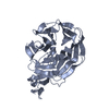
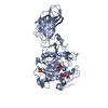
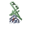
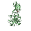
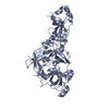
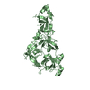
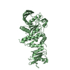
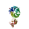
| Title | Tandem YVTN beta-propeller and PKD domains from an archaeal surface layer protein | ||||||
 Components Components | Surface layer protein | ||||||
 Keywords Keywords | PROTEIN BINDING / surface layer protein / SLP / S-layer / 7-bladed beta-propeller / PKD superfamily | ||||||
| Function / homology |  Function and homology information Function and homology information | ||||||
| Biological species |  Methanosarcina mazei (archaea) Methanosarcina mazei (archaea) | ||||||
| Method | X-RAY DIFFRACTION / SYNCHROTRON / MAD / Resolution: 2.4 Å | ||||||
 Authors Authors | Jing, H. / Takagi, J. / Liu, J.-H. / Lindgren, S. / Zhang, R.-G. / Joachimiak, A. / Wang, J.-H. / Springer, T.A. | ||||||
 Citation Citation | Journal: Structure / Year: 2002 Title: Archaeal Surface Layer Proteins Contain beta Propeller, PKD, and beta Helix Domains and Are Related to Metazoan Cell Surface Proteins. Authors: Jing, H. / Takagi, J. / Liu, J. / Lindgren, S. / Zhang, R. / Joachimiak, A. / Wang, J. / Springer, T. #1: Journal: Nat.Struct.Biol. / Year: 2001Title: Implications for familial hypercholesterolemia from structure of the LDL receptor YWTD-EGF doman pair Authors: Jeon, H. / Meng, W. / Takagi, J. / Eck, M.J. / Springer, T.A. / Blacklow, S.C. #2: Journal: Embo J. / Year: 1999Title: The structure of a PKD domain from polycystin-1: implications for polycystic kidney disease Authors: Bycroft, M. / Bateman, A. / Clarke, J. / Hamill, S.J. / Sandford, R. / Thomas, R.L. / Chothia, C. #3: Journal: Arch.Microbiol. / Year: 1998Title: Structure, organization, and expression of genes coding for envelope components in the archaeon Methanosarcina mazei S-6 Authors: Mayerhofer, L.E. / Conway de Macario, E. / Yao, R. / Macario, A.J.L. #4: Journal: To be PublishedTitle: The genome of Methanosarcina acetivorans reveals extensive metabolic and physiological diversity Authors: Galagan, J.E. / Nusbaum, C. / Roy, A. / Endrizzi, M.G. / Macdonald, P. / FitzHugh, W. / Calvo, S. / Engels, R. / Smirnov, S. #5: Journal: J.Mol.Biol. / Year: 1998Title: An extracellular beta-propeller module predicted in lipoprotein and scavenger receptors, tyrosine kinases, epidermal growth factor precursor, and extracellular matrix components Authors: Springer, T.A. | ||||||
| History |
| ||||||
| Remark 300 | BIOMOLECULE: 1 THIS ENTRY CONTAINS THE CRYSTALLOGRAPHIC ASYMMETRIC UNIT WHICH CONSISTS OF 4 ... BIOMOLECULE: 1 THIS ENTRY CONTAINS THE CRYSTALLOGRAPHIC ASYMMETRIC UNIT WHICH CONSISTS OF 4 CHAIN(S). SEE REMARK 350 FOR INFORMATION ON GENERATING THE BIOLOGICAL MOLECULE(S). THE WHOLE SURFACE LAYER PROTEIN MOLECULE HAS A DOMAIN ORGANIZATION OF YVTN-PKD-YVTN-(PKD)11. THE COORDINATES OF ANY ONE OF THE 4 MOLECULES IN THE ASYMMETRIC UNIT COULD CORRESPOND TO THE BIOLOGICAL CONFORMATION OF THE N-TERMINAL YVTN-PKD DOMAIN PAIR. |
- Structure visualization
| Structure viewer | Molecule: MolmilJmol/JSmol |
|---|
- Downloads & links
-Download
| PDBx/mmCIF format | 1l0q.cif.gz | 313.2 KB | Display | PDBx/mmCIF format |
|---|---|---|---|---|
| PDB format | pdb1l0q.ent.gz | 264.4 KB | Display | PDB format |
| PDBx/mmJSON format | 1l0q.json.gz | Tree view | PDBx/mmJSON format | |
| Others |  Other downloads Other downloads |
-Validation report
| Arichive directory | https://data.pdbj.org/pub/pdb/validation_reports/l0/1l0qftp://data.pdbj.org/pub/pdb/validation_reports/l0/1l0q | HTTPS FTP |
|---|
-Related structure data
| Related structure data | |
|---|---|
| Similar structure data |


-Links
 PDBj
PDBj- Assembly
| Deposited unit | 
| ||||||||
|---|---|---|---|---|---|---|---|---|---|
| 1 | 
| ||||||||
| 2 | 
| ||||||||
| 3 | 
| ||||||||
| 4 | 
| ||||||||
| Unit cell |
|
-Components
| #1: Protein | Mass: 40796.559 Da / Num. of mol.: 4 / Fragment: YVTN beta-propeller domain 1 and PKD domain 1 Source method: isolated from a genetically manipulated source Source: (gene. exp.) Methanosarcina mazei (archaea) / Strain: S-6 / Plasmid: pQE-60 / Production host:  Escherichia coli (E. coli) / Strain (production host): M15 / References: UniProt: Q50245 Escherichia coli (E. coli) / Strain (production host): M15 / References: UniProt: Q50245#2: Water | ChemComp-HOH / | Water Mass: 18.015 Da / Num. of mol.: 1364 / Source method: isolated from a natural source / Formula: H2O Mass: 18.015 Da / Num. of mol.: 1364 / Source method: isolated from a natural source / Formula: H2O |
|---|
-Experimental details
-Experiment
| Experiment | Method: X-RAY DIFFRACTION / Number of used crystals: 1 |
|---|
- Sample preparation
| Crystal | Density Matthews: 2.16 Å3/Da / Density % sol: 42.7 % | |||||||||||||||||||||||||||||||||||
|---|---|---|---|---|---|---|---|---|---|---|---|---|---|---|---|---|---|---|---|---|---|---|---|---|---|---|---|---|---|---|---|---|---|---|---|---|
| Crystal grow | Temperature: 298 K / Method: vapor diffusion, hanging drop / pH: 3.8 Details: PEG-8000, potassium chloride, sodium acetate, pH 3.8, VAPOR DIFFUSION, HANGING DROP, temperature 298.0K | |||||||||||||||||||||||||||||||||||
| Crystal grow | *PLUS | |||||||||||||||||||||||||||||||||||
| Components of the solutions | *PLUS
|
-Data collection
| Diffraction | Mean temperature: 200 K | ||||||||||||
|---|---|---|---|---|---|---|---|---|---|---|---|---|---|
| Diffraction source | Source: SYNCHROTRON / Site: APS  / Beamline: 19-ID / Wavelength: 0.979067,0.979221,0.939270 / Beamline: 19-ID / Wavelength: 0.979067,0.979221,0.939270 | ||||||||||||
| Detector | Type: SBC-2 / Detector: CCD / Date: Nov 24, 1999 / Details: mirrors | ||||||||||||
| Radiation | Monochromator: SAGITALLY FOCUSED Si(111) / Protocol: MAD / Monochromatic (M) / Laue (L): M / Scattering type: x-ray | ||||||||||||
| Radiation wavelength |
| ||||||||||||
| Reflection | Resolution: 2.4→100 Å / Num. all: 52881 / Num. obs: 52881 / % possible obs: 93.7 % / Observed criterion σ(F): 0 / Observed criterion σ(I): 0 / Redundancy: 6 % / Biso Wilson estimate: 30.36 Å2 / Rmerge(I) obs: 0.109 / Net I/σ(I): 14.6 | ||||||||||||
| Reflection shell | Resolution: 2.4→2.49 Å / Rmerge(I) obs: 0.434 / Mean I/σ(I) obs: 3.1 / Num. unique all: 4965 / % possible all: 89 | ||||||||||||
| Reflection | *PLUS Lowest resolution: 100 Å / Num. measured all: 318719 / Rmerge(I) obs: 0.109 | ||||||||||||
| Reflection shell | *PLUS % possible obs: 89 % / Rmerge(I) obs: 0.434 |
- Processing
| Software |
| ||||||||||||||||||||||||||||||||||||
|---|---|---|---|---|---|---|---|---|---|---|---|---|---|---|---|---|---|---|---|---|---|---|---|---|---|---|---|---|---|---|---|---|---|---|---|---|---|
| Refinement | Method to determine structure: MAD / Resolution: 2.4→20 Å / Data cutoff high rms absF: 1000 / Isotropic thermal model: isotropic / Cross valid method: THROUGHOUT / σ(F): 0 / Stereochemistry target values: Engh & Huber
| ||||||||||||||||||||||||||||||||||||
| Solvent computation | Bsol: 40.168 Å2 / ksol: 0.323 e/Å3 | ||||||||||||||||||||||||||||||||||||
| Displacement parameters | Biso mean: 23.4 Å2 | ||||||||||||||||||||||||||||||||||||
| Refine analyze |
| ||||||||||||||||||||||||||||||||||||
| Refinement step | Cycle: LAST / Resolution: 2.4→20 Å
| ||||||||||||||||||||||||||||||||||||
| Refine LS restraints |
| ||||||||||||||||||||||||||||||||||||
| LS refinement shell | Resolution: 2.4→2.49 Å / Total num. of bins used: 10
| ||||||||||||||||||||||||||||||||||||
| Refinement | *PLUS Lowest resolution: 20 Å / % reflection Rfree: 5 % / Rfactor obs: 0.217 / Rfactor Rfree: 0.263 / Rfactor Rwork: 0.214 | ||||||||||||||||||||||||||||||||||||
| Solvent computation | *PLUS | ||||||||||||||||||||||||||||||||||||
| Displacement parameters | *PLUS | ||||||||||||||||||||||||||||||||||||
| Refine LS restraints | *PLUS
| ||||||||||||||||||||||||||||||||||||
| LS refinement shell | *PLUS Rfactor Rfree: 0.3466 / Rfactor Rwork: 0.2897 |
