 Movie
Movie Controller

[English] 日本語
 Yorodumi
- PDB-1kcd: Endopolygalacturonase I from Stereum purpureum complexed with two... -
+ Open data

- Basic information
| Entry | Database: PDB / ID: 1kcd | ||||||||||||
|---|---|---|---|---|---|---|---|---|---|---|---|---|---|
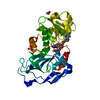
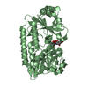
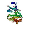
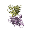
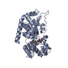
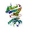
| Title | Endopolygalacturonase I from Stereum purpureum complexed with two galacturonate at 1.15 A resolution. | ||||||||||||
 Components Components | ENDOPOLYGALACTURONASE Polygalacturonase Polygalacturonase | ||||||||||||
 Keywords Keywords | HYDROLASE / BETA HELICAL STRUCTURE / GLYCOSIDE HYDROLASE / SILVER-LEAF INDUCING SUBSTANCE | ||||||||||||
| Function / homology |  Function and homology informationendo-polygalacturonase / polygalacturonase activity / pectin catabolic process / cell wall organization Function and homology informationendo-polygalacturonase / polygalacturonase activity / pectin catabolic process / cell wall organizationSimilarity search - Function | ||||||||||||
| Biological species |  Chondrostereum purpureum (fungus) Chondrostereum purpureum (fungus) | ||||||||||||
| Method | X-RAY DIFFRACTION / SYNCHROTRON / MOLECULAR REPLACEMENT / Resolution: 1.15 Å | ||||||||||||
 Authors Authors | Shimizu, T. / Nakatsu, T. / Miyairi, K. / Okuno, T. / Kato, H. | ||||||||||||
 Citation Citation | Journal: Biochemistry / Year: 2002 Title: Active-site architecture of endopolygalacturonase I from Stereum purpureum revealed by crystal structures in native and ligand-bound forms at atomic resolution. Authors: Shimizu, T. / Nakatsu, T. / Miyairi, K. / Okuno, T. / Kato, H. #1: Journal: Acta Crystallogr.,Sect.D / Year: 2001Title: Crystallization and preliminary X-ray study of endopolygalacturonase from the pathogenic fungus Stereum purpureum Authors: Shimizu, T. / Nakatsu, T. / Miyairi, K. / Okuno, T. / Kato, H. #2: Journal: Eur.J.Biochem. / Year: 2000Title: Determination of glycosylation sites, disulfide bridges, and the C-terminus of Stereum purpureum mature endopolygalacturonase I by electrospray ionization mass spectrometry Authors: Shimizu, T. / Miyairi, K. / Okuno, T. #3: Journal: BIOSCI.BIOTECHNOL.BIOCHEM. / Year: 1998Title: Isolation, characterization, and sugar chain structure of endoPG Ia, Ib and Ic from Stereum purpureum Authors: Hasui, Y. / Fukui, Y. / Kikuchi, J. / Kato, N. / Miyairi, K. / Okuno, T. | ||||||||||||
| History |
|
- Structure visualization
| Structure viewer | Molecule: MolmilJmol/JSmol |
|---|
- Downloads & links
-Download
| PDBx/mmCIF format | 1kcd.cif.gz | 160.2 KB | Display | PDBx/mmCIF format |
|---|---|---|---|---|
| PDB format | pdb1kcd.ent.gz | 124.3 KB | Display | PDB format |
| PDBx/mmJSON format | 1kcd.json.gz | Tree view | PDBx/mmJSON format | |
| Others |  Other downloads Other downloads |
-Validation report
| Arichive directory | https://data.pdbj.org/pub/pdb/validation_reports/kc/1kcdftp://data.pdbj.org/pub/pdb/validation_reports/kc/1kcd | HTTPS FTP |
|---|
-Related structure data
| Related structure data |  1k5cSC  1kccC S: Starting model for refinement C: citing same article ( |
|---|---|
| Similar structure data |


-Links
 PDBj
PDBj- Assembly
| Deposited unit | 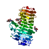
| ||||||||
|---|---|---|---|---|---|---|---|---|---|
| 1 |
| ||||||||
| Unit cell |
|
-Components
-Protein , 1 types, 1 molecules A
| #1: Protein | Polygalacturonase Mass: 34652.363 Da / Num. of mol.: 1 / Fragment: residues 1-335 / Source method: isolated from a natural source / Source: (natural) Chondrostereum purpureum (fungus) / Strain: ASP-4B / References: UniProt: P79074, endo-polygalacturonase |
|---|
-Sugars , 3 types, 4 molecules 


| #2: Sugar | ChemComp-GTK / D-Galacturonic acid Type: D-saccharide, beta linking / Mass: 194.139 Da / Num. of mol.: 1 Type: D-saccharide, beta linking / Mass: 194.139 Da / Num. of mol.: 1Source method: isolated from a genetically manipulated source Formula: C6H10O7 |
|---|---|
| #3: Sugar | ChemComp-GTR / D-Galacturonic acid Type: D-saccharide, beta linking / Mass: 194.139 Da / Num. of mol.: 1 Type: D-saccharide, beta linking / Mass: 194.139 Da / Num. of mol.: 1Source method: isolated from a genetically manipulated source Formula: C6H10O7 |
| #4: Sugar | N-Acetylglucosamine Type: D-saccharide, beta linking / Mass: 221.208 Da / Num. of mol.: 2 Type: D-saccharide, beta linking / Mass: 221.208 Da / Num. of mol.: 2Source method: isolated from a genetically manipulated source Formula: C8H15NO6 |
-Non-polymers , 2 types, 521 molecules 

| #5: Chemical | ChemComp-CL / Chloride Mass: 35.453 Da / Num. of mol.: 1 / Source method: obtained synthetically / Formula: Cl Mass: 35.453 Da / Num. of mol.: 1 / Source method: obtained synthetically / Formula: Cl |
|---|---|
| #6: Water | ChemComp-HOH / WaterMass: 18.015 Da / Num. of mol.: 520 / Source method: isolated from a natural source / Formula: H2O |
-Experimental details
-Experiment
| Experiment | Method: X-RAY DIFFRACTION / Number of used crystals: 1 |
|---|
- Sample preparation
| Crystal | Density Matthews: 1.66 Å3/Da / Density % sol: 45.22 % | ||||||||||||||||||||||||||||||||||||||||||
|---|---|---|---|---|---|---|---|---|---|---|---|---|---|---|---|---|---|---|---|---|---|---|---|---|---|---|---|---|---|---|---|---|---|---|---|---|---|---|---|---|---|---|---|
| Crystal grow | Temperature: 293 K / Method: vapor diffusion, hanging drop / pH: 5 Details: PEG 4000, pH 5.0, VAPOR DIFFUSION, HANGING DROP, temperature 293K | ||||||||||||||||||||||||||||||||||||||||||
| Crystal grow | *PLUS Details: Shimizu, T., (2001) Acta Crystallogr., Sect.D, 57, 1171. | ||||||||||||||||||||||||||||||||||||||||||
| Components of the solutions | *PLUS
|
-Data collection
| Diffraction | Mean temperature: 90 K |
|---|---|
| Diffraction source | Source: SYNCHROTRON / Site: SPring-8  / Beamline: BL44B2 / Wavelength: 0.6 Å / Beamline: BL44B2 / Wavelength: 0.6 Å |
| Detector | Type: MAR CCD 165 mm / Detector: CCD / Date: Sep 27, 2001 |
| Radiation | Protocol: SINGLE WAVELENGTH / Monochromatic (M) / Laue (L): M / Scattering type: x-ray |
| Radiation wavelength | Wavelength: 0.6 Å / Relative weight: 1 |
| Reflection | Resolution: 1.15→12.1 Å / Num. all: 104270 / Num. obs: 104270 / % possible obs: 97.3 % / Observed criterion σ(F): 1 / Observed criterion σ(I): 1 / Redundancy: 2.2 % / Biso Wilson estimate: 5.6 Å2 / Rsym value: 0.071 / Net I/σ(I): 6.5 |
| Reflection shell | Resolution: 1.15→1.21 Å / Redundancy: 2.2 % / Mean I/σ(I) obs: 2.8 / Num. unique all: 15075 / Rsym value: 0.251 / % possible all: 96.2 |
| Reflection | *PLUS Num. measured all: 230114 / Rmerge(I) obs: 0.071 |
| Reflection shell | *PLUS % possible obs: 96.2 % / Rmerge(I) obs: 0.251 |
- Processing
| Software |
| |||||||||||||||||||||||||||||||||
|---|---|---|---|---|---|---|---|---|---|---|---|---|---|---|---|---|---|---|---|---|---|---|---|---|---|---|---|---|---|---|---|---|---|---|
| Refinement | Method to determine structure: MOLECULAR REPLACEMENT Starting model: PDB ENTRY 1K5C Resolution: 1.15→10 Å / Num. parameters: 27121 / Num. restraintsaints: 32146 / Cross valid method: FREE R / σ(F): 0 / Stereochemistry target values: ENGH AND HUBER
| |||||||||||||||||||||||||||||||||
| Refine analyze | Num. disordered residues: 11 / Occupancy sum hydrogen: 0 / Occupancy sum non hydrogen: 2925.52 | |||||||||||||||||||||||||||||||||
| Refinement step | Cycle: LAST / Resolution: 1.15→10 Å
| |||||||||||||||||||||||||||||||||
| Refine LS restraints |
| |||||||||||||||||||||||||||||||||
| Software | *PLUS Name: SHELXL / Version: 97 / Classification: refinement | |||||||||||||||||||||||||||||||||
| Refinement | *PLUS Lowest resolution: 10 Å / Rfactor all: 0.135 / Rfactor obs: 0.126 / Rfactor Rfree: 0.164 / Rfactor Rwork: 0.126 | |||||||||||||||||||||||||||||||||
| Solvent computation | *PLUS | |||||||||||||||||||||||||||||||||
| Displacement parameters | *PLUS | |||||||||||||||||||||||||||||||||
| Refine LS restraints | *PLUS
|
