 Movie
Movie Controller


+ Open data

- Basic information
| Entry | Database: PDB / ID: 1k9o | |||||||||
|---|---|---|---|---|---|---|---|---|---|---|
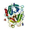
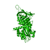
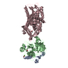
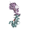
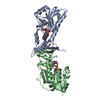
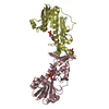
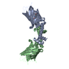
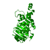
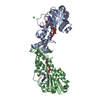
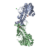
| Title | CRYSTAL STRUCTURE OF MICHAELIS SERPIN-TRYPSIN COMPLEX | |||||||||
 Components Components |
| |||||||||
 Keywords Keywords | Hydrolase/Hydrolase Inhibitor / Michaelis serpin-protease complex inhibitory triad / Hydrolase-Hydrolase Inhibitor COMPLEX | |||||||||
| Function / homology |  Function and homology information Function and homology informationAntimicrobial peptides / Alpha-defensins / Activation of Matrix Metalloproteinases / trypsin / Neutrophil degranulation / digestion / collagen catabolic process / response to nutrient / response to bacterium / serine-type endopeptidase inhibitor activity ...Antimicrobial peptides / Alpha-defensins / Activation of Matrix Metalloproteinases / trypsin / Neutrophil degranulation / digestion / collagen catabolic process / response to nutrient / response to bacterium / serine-type endopeptidase inhibitor activity / serine-type endopeptidase activity / calcium ion binding / proteolysis / extracellular space / extracellular region Similarity search - Function | |||||||||
| Biological species |  Manduca sexta (tobacco hornworm) Manduca sexta (tobacco hornworm) | |||||||||
| Method |  X-RAY DIFFRACTION / SYNCHROTRON / MOLECULAR REPLACEMENT / Resolution: 2.3 Å X-RAY DIFFRACTION / SYNCHROTRON / MOLECULAR REPLACEMENT / Resolution: 2.3 Å | |||||||||
 Authors Authors | Ye, S. / Cech, A.L. / Belmares, R. / Bergstrom, R.C. / Tong, Y. / Corey, D.R. / Kanost, M.R. / Goldsmith, E.J. | |||||||||
 Citation Citation | Journal: Nat.Struct.Biol. / Year: 2001 Title: The structure of a Michaelis serpin-protease complex. Authors: Ye, S. / Cech, A.L. / Belmares, R. / Bergstrom, R.C. / Tong, Y. / Corey, D.R. / Kanost, M.R. / Goldsmith, E.J. | |||||||||
| History |
|
- Structure visualization
| Structure viewer | Molecule: MolmilJmol/JSmol |
|---|
- Downloads & links
-Download
| PDBx/mmCIF format | 1k9o.cif.gz | 132.4 KB | Display | PDBx/mmCIF format |
|---|---|---|---|---|
| PDB format | pdb1k9o.ent.gz | 101.2 KB | Display | PDB format |
| PDBx/mmJSON format | 1k9o.json.gz | Tree view | PDBx/mmJSON format | |
| Others |  Other downloads Other downloads |
-Validation report
| Summary document | 1k9o_validation.pdf.gz | 433.1 KB | Display | wwPDB validaton report |
|---|---|---|---|---|
| Full document | 1k9o_full_validation.pdf.gz | 451.8 KB | Display | |
| Data in XML | 1k9o_validation.xml.gz | 25.8 KB | Display | |
| Data in CIF | 1k9o_validation.cif.gz | 36.2 KB | Display | |
| Arichive directory | https://data.pdbj.org/pub/pdb/validation_reports/k9/1k9oftp://data.pdbj.org/pub/pdb/validation_reports/k9/1k9o | HTTPS FTP |
-Related structure data


-Links
 PDBj
PDBj


- Assembly
| Deposited unit | 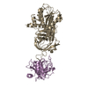
| |||||||||
|---|---|---|---|---|---|---|---|---|---|---|
| 1 |
| |||||||||
| Unit cell |
| |||||||||
| Components on special symmetry positions |
|
-Components
| #1: Protein | Mass: 42016.988 Da / Num. of mol.: 1 / Mutation: A353K Source method: isolated from a genetically manipulated source Source: (gene. exp.) Manduca sexta (tobacco hornworm) / Plasmid: H6PQE-60 / Production host:  |
|---|---|
| #2: Protein | Mass: 23798.838 Da / Num. of mol.: 1 / Mutation: S195A Source method: isolated from a genetically manipulated source Source: (gene. exp.)  |
| #3: Water | ChemComp-HOH /  Mass: 18.015 Da / Num. of mol.: 182 / Source method: isolated from a natural source / Formula: H2O Mass: 18.015 Da / Num. of mol.: 182 / Source method: isolated from a natural source / Formula: H2O |
-Experimental details
-Experiment
| Experiment | Method: X-RAY DIFFRACTION / Number of used crystals: 1 |
|---|
- Sample preparation
| Crystal | Density Matthews: 2.67 Å3/Da / Density % sol: 53.86 % | ||||||||||||||||||||||||||||||||||||||||||||||||||||||||||||
|---|---|---|---|---|---|---|---|---|---|---|---|---|---|---|---|---|---|---|---|---|---|---|---|---|---|---|---|---|---|---|---|---|---|---|---|---|---|---|---|---|---|---|---|---|---|---|---|---|---|---|---|---|---|---|---|---|---|---|---|---|---|
| Crystal grow | Temperature: 298 K / Method: vapor diffusion, hanging drop / pH: 6.5 Details: PEG 3350, DTT, POTASSIUM PHOSPHATE, SODIUM PHOSPHATE, 2-PROPANOL, AMMONIUM CHLORIDE, pH 6.5, VAPOR DIFFUSION, HANGING DROP, temperature 298K | ||||||||||||||||||||||||||||||||||||||||||||||||||||||||||||
| Crystal grow | *PLUS PH range low: 6.5 / PH range high: 6 | ||||||||||||||||||||||||||||||||||||||||||||||||||||||||||||
| Components of the solutions | *PLUS
|
-Data collection
| Diffraction | Mean temperature: 100 K |
|---|---|
| Diffraction source | Source: SYNCHROTRON / Site: APS  / Beamline: 19-ID / Wavelength: 1.0332 Å / Beamline: 19-ID / Wavelength: 1.0332 Å |
| Detector | Detector: CCD / Date: Dec 10, 1999 / Details: MIRRORS |
| Radiation | Protocol: SINGLE WAVELENGTH / Monochromatic (M) / Laue (L): M / Scattering type: x-ray |
| Radiation wavelength | Wavelength: 1.0332 Å / Relative weight: 1 |
| Reflection | Resolution: 2.3→20 Å / Num. obs: 30999 / % possible obs: 98.4 % / Redundancy: 6 % / Rmerge(I) obs: 0.091 / Net I/σ(I): 11.3 |
| Reflection shell | Resolution: 2.3→2.38 Å / Redundancy: 6 % / Rmerge(I) obs: 0.391 / Mean I/σ(I) obs: 1.76 / Num. unique all: 30999 / % possible all: 95.6 |
| Reflection | *PLUS Num. measured all: 186342 |
| Reflection shell | *PLUS % possible obs: 95.6 % |
- Processing
| Software |
| ||||||||||||||||||||
|---|---|---|---|---|---|---|---|---|---|---|---|---|---|---|---|---|---|---|---|---|---|
| Refinement | Method to determine structure: MOLECULAR REPLACEMENT Starting model: PDB ENTRIES 1SEK AND 1DPO Resolution: 2.3→20 Å / Cross valid method: THROUGHOUT / σ(F): 2
| ||||||||||||||||||||
| Refinement step | Cycle: LAST / Resolution: 2.3→20 Å
| ||||||||||||||||||||
| Refine LS restraints |
| ||||||||||||||||||||
| Software | *PLUS Name: CNS / Version: 1 / Classification: refinement | ||||||||||||||||||||
| Refinement | *PLUS Highest resolution: 2.3 Å / Lowest resolution: 20 Å / σ(F): 2 / Rfactor obs: 0.158 | ||||||||||||||||||||
| Solvent computation | *PLUS | ||||||||||||||||||||
| Displacement parameters | *PLUS | ||||||||||||||||||||
| Refine LS restraints | *PLUS
|
