 Movie
Movie Controller

[English] 日本語
 Yorodumi
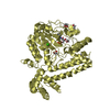
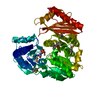
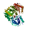
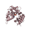
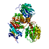
- PDB-1k2y: Crystal Structure of Phosphomannomutase/Phosphoglucomutase S108A ... -
+ Open data

- Basic information
| Entry | Database: PDB / ID: 1k2y | ||||||
|---|---|---|---|---|---|---|---|
| Title | Crystal Structure of Phosphomannomutase/Phosphoglucomutase S108A mutant from P. aeruginosa | ||||||
 Components Components | phosphomannomutase | ||||||
 Keywords Keywords | ISOMERASE / ALPHA/BETA PROTEIN / ACTIVE-SITE MUTANT / ENZYME-LIGAND COMPLEX | ||||||
| Function / homology |  Function and homology information Function and homology informationphosphomannomutase / phosphoglucomutase (alpha-D-glucose-1,6-bisphosphate-dependent) / phosphoglucomutase activity / phosphomannomutase activity / alginic acid biosynthetic process / O antigen biosynthetic process / GDP-mannose biosynthetic process / lipopolysaccharide core region biosynthetic process / magnesium ion binding Similarity search - Function | ||||||
| Biological species |   Pseudomonas aeruginosa (bacteria) Pseudomonas aeruginosa (bacteria) | ||||||
| Method |  X-RAY DIFFRACTION / refinement of wild-type structure / Resolution: 1.75 Å X-RAY DIFFRACTION / refinement of wild-type structure / Resolution: 1.75 Å | ||||||
 Authors Authors | Regni, C. / Tipton, P.A. / Beamer, L.J. | ||||||
 Citation Citation | Journal: Structure / Year: 2002 Title: Crystal structure of PMM/PGM: an enzyme in the biosynthetic pathway of P. aeruginosa virulence factors. Authors: Regni, C. / Tipton, P.A. / Beamer, L.J. | ||||||
| History |
|
- Structure visualization
| Structure viewer | Molecule: MolmilJmol/JSmol |
|---|
- Downloads & links
-Download
| PDBx/mmCIF format | 1k2y.cif.gz | 108.9 KB | Display | PDBx/mmCIF format |
|---|---|---|---|---|
| PDB format | pdb1k2y.ent.gz | 82.3 KB | Display | PDB format |
| PDBx/mmJSON format | 1k2y.json.gz | Tree view | PDBx/mmJSON format | |
| Others |  Other downloads Other downloads |
-Validation report
| Arichive directory | https://data.pdbj.org/pub/pdb/validation_reports/k2/1k2yftp://data.pdbj.org/pub/pdb/validation_reports/k2/1k2y | HTTPS FTP |
|---|
-Related structure data
| Related structure data |  1k35SC S: Starting model for refinement C: citing same article ( |
|---|---|
| Similar structure data |


-Links
 PDBj
PDBj
- Assembly
| Deposited unit | 
| ||||||||
|---|---|---|---|---|---|---|---|---|---|
| 1 |
| ||||||||
| Unit cell |
|
-Components
| #1: Protein | Mass: 50334.352 Da / Num. of mol.: 1 / Mutation: S108A Source method: isolated from a genetically manipulated source Source: (gene. exp.) Pseudomonas aeruginosa (bacteria) / Gene: AlgC / Plasmid: pet3-A / Species (production host): Escherichia coli / Production host: |
|---|---|
| #2: Chemical | ChemComp-ZN /   Mass: 65.409 Da / Num. of mol.: 1 / Source method: obtained synthetically / Formula: Zn Mass: 65.409 Da / Num. of mol.: 1 / Source method: obtained synthetically / Formula: Zn |
| #3: Chemical | ChemComp-TLA /   Mass: 150.087 Da / Num. of mol.: 1 / Source method: obtained synthetically / Formula: C4H6O6 Mass: 150.087 Da / Num. of mol.: 1 / Source method: obtained synthetically / Formula: C4H6O6 |
| #4: Water | ChemComp-HOH /  Mass: 18.015 Da / Num. of mol.: 404 / Source method: isolated from a natural source / Formula: H2O Mass: 18.015 Da / Num. of mol.: 404 / Source method: isolated from a natural source / Formula: H2O |
-Experimental details
-Experiment
| Experiment | Method: X-RAY DIFFRACTION / Number of used crystals: 1 |
|---|
- Sample preparation
| Crystal | Density Matthews: 2.25 Å3/Da / Density % sol: 44.8 % | ||||||||||||||||||||||||||||||
|---|---|---|---|---|---|---|---|---|---|---|---|---|---|---|---|---|---|---|---|---|---|---|---|---|---|---|---|---|---|---|---|
| Crystal grow | Temperature: 298 K / Method: vapor diffusion, hanging drop / pH: 7 Details: Na,K tartrate, MOPS, pH 7.0, VAPOR DIFFUSION, HANGING DROP, temperature 298K | ||||||||||||||||||||||||||||||
| Crystal grow | *PLUS Details: Regni, C.A., (2000) Acta Crystallogr, D56, 761. | ||||||||||||||||||||||||||||||
| Components of the solutions | *PLUS
|
-Data collection
| Diffraction | Mean temperature: 100 K |
|---|---|
| Diffraction source | Source: ROTATING ANODE / Type: RIGAKU RU200 / Wavelength: 1.5418 |
| Detector | Type: RIGAKU RAXIS IV / Detector: IMAGE PLATE / Details: Osmic confocal |
| Radiation | Monochromator: OSMIC / Protocol: SINGLE WAVELENGTH / Monochromatic (M) / Laue (L): M / Scattering type: x-ray |
| Radiation wavelength | Wavelength: 1.5418 Å / Relative weight: 1 |
| Reflection | Resolution: 1.75→40 Å / Num. all: 49105 / Num. obs: 49105 / % possible obs: 97.4 % / Observed criterion σ(F): 0 / Observed criterion σ(I): 0 / Redundancy: 4.9 % / Biso Wilson estimate: 33.22 Å2 / Rmerge(I) obs: 0.053 / Net I/σ(I): 28.5 |
| Reflection shell | Resolution: 1.75→1.81 Å / Rmerge(I) obs: 0.363 / Mean I/σ(I) obs: 2.3 / % possible all: 98.8 |
| Reflection | *PLUS Lowest resolution: 40 Å / Num. measured all: 240203 |
| Reflection shell | *PLUS % possible obs: 98.8 % |
- Processing
| Software |
| ||||||||||||||||||||||||||||||||||||||||||||||||||||
|---|---|---|---|---|---|---|---|---|---|---|---|---|---|---|---|---|---|---|---|---|---|---|---|---|---|---|---|---|---|---|---|---|---|---|---|---|---|---|---|---|---|---|---|---|---|---|---|---|---|---|---|---|---|
| Refinement | Method to determine structure: refinement of wild-type structure Starting model: Native protein, pdb entry 1k35 Resolution: 1.75→40 Å / SU B: 2.9191 / SU ML: 0.09446 / Cross valid method: THROUGHOUT / σ(F): 0 / ESU R: 0.10699 / ESU R Free: 0.10087 / Stereochemistry target values: Engh & Huber
| ||||||||||||||||||||||||||||||||||||||||||||||||||||
| Displacement parameters | Biso mean: 28.905 Å2
| ||||||||||||||||||||||||||||||||||||||||||||||||||||
| Refinement step | Cycle: LAST / Resolution: 1.75→40 Å
| ||||||||||||||||||||||||||||||||||||||||||||||||||||
| Refine LS restraints |
| ||||||||||||||||||||||||||||||||||||||||||||||||||||
| Software | *PLUS Name: REFMAC / Classification: refinement | ||||||||||||||||||||||||||||||||||||||||||||||||||||
| Refinement | *PLUS σ(F): 0 / % reflection Rfree: 5 % / Rfactor all: 0.17 / Rfactor Rfree: 0.196 / Rfactor Rwork: 0.169 | ||||||||||||||||||||||||||||||||||||||||||||||||||||
| Solvent computation | *PLUS | ||||||||||||||||||||||||||||||||||||||||||||||||||||
| Displacement parameters | *PLUS | ||||||||||||||||||||||||||||||||||||||||||||||||||||
| Refine LS restraints | *PLUS Type: p_angle_deg / Dev ideal: 1.6 |
