 Movie
Movie Controller


+ Open data

- Basic information
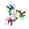
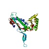
| Entry | Database: PDB / ID: 1js0 | ||||||
|---|---|---|---|---|---|---|---|
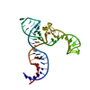
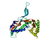
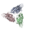
| Title | Crystal Structure of 3D Domain-swapped RNase A Minor Trimer | ||||||
 Components Components | RIBONUCLEASE A | ||||||
 Keywords Keywords | HYDROLASE / 3D domain swapping / RNase A | ||||||
| Function / homology |  Function and homology information Function and homology informationpancreatic ribonuclease / ribonuclease A activity / RNA nuclease activity / nucleic acid binding / defense response to Gram-positive bacterium / hydrolase activity / extracellular region Similarity search - Function | ||||||
| Biological species |  | ||||||
| Method |  X-RAY DIFFRACTION / SYNCHROTRON / MOLECULAR REPLACEMENT / Resolution: 2.2 Å X-RAY DIFFRACTION / SYNCHROTRON / MOLECULAR REPLACEMENT / Resolution: 2.2 Å | ||||||
 Authors Authors | Liu, Y. / Gotte, G. / Libonati, M. / Eisenberg, D. | ||||||
 Citation Citation | Journal: Protein Sci. / Year: 2002 Title: Structures of the two 3D domain-swapped RNase A trimers. Authors: Liu, Y. / Gotte, G. / Libonati, M. / Eisenberg, D. #1: Journal: Proc.Natl.Acad.Sci.USA / Year: 1998Title: The Crystal Structure of a 3D Domain-swapped Dimer of RNase A at a 2.1 A Resolution Authors: Liu, Y. / Hart, P.J. / Schlunegger, M.P. / Eisenberg, D. #2: Journal: Nat.Struct.Biol. / Year: 2001Title: A domain-swapped RNase A dimer with implications for amyloid formation Authors: Liu, Y. / Gotte, G. / Libonati, M. / Eisenberg, D. | ||||||
| History |
|
- Structure visualization
| Structure viewer | Molecule: MolmilJmol/JSmol |
|---|
- Downloads & links
-Download
| PDBx/mmCIF format | 1js0.cif.gz | 88.2 KB | Display | PDBx/mmCIF format |
|---|---|---|---|---|
| PDB format | pdb1js0.ent.gz | 68.3 KB | Display | PDB format |
| PDBx/mmJSON format | 1js0.json.gz | Tree view | PDBx/mmJSON format | |
| Others |  Other downloads Other downloads |
-Validation report
| Arichive directory | https://data.pdbj.org/pub/pdb/validation_reports/js/1js0ftp://data.pdbj.org/pub/pdb/validation_reports/js/1js0 | HTTPS FTP |
|---|
-Related structure data
| Related structure data | 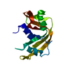 1rtbS S: Starting model for refinement |
|---|---|
| Similar structure data |


-Links
 PDBj
PDBj

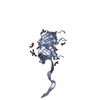
- Assembly
| Deposited unit | 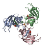
| ||||||||
|---|---|---|---|---|---|---|---|---|---|
| 1 |
| ||||||||
| Unit cell |
| ||||||||
| Components on special symmetry positions |
|
-Components
| #1: Protein | Mass: 13708.326 Da / Num. of mol.: 3 / Source method: isolated from a natural source / Source: (natural) #2: Chemical | ChemComp-SO4 /   Mass: 96.063 Da / Num. of mol.: 4 / Source method: obtained synthetically / Formula: SO4 Mass: 96.063 Da / Num. of mol.: 4 / Source method: obtained synthetically / Formula: SO4#3: Water | ChemComp-HOH / |  Mass: 18.015 Da / Num. of mol.: 267 / Source method: isolated from a natural source / Formula: H2O Mass: 18.015 Da / Num. of mol.: 267 / Source method: isolated from a natural source / Formula: H2OHas protein modification | Y | |
|---|
-Experimental details
-Experiment
| Experiment | Method: X-RAY DIFFRACTION / Number of used crystals: 1 |
|---|
- Sample preparation
| Crystal | Density Matthews: 2.28 Å3/Da / Density % sol: 46.13 % | ||||||||||||||||||
|---|---|---|---|---|---|---|---|---|---|---|---|---|---|---|---|---|---|---|---|
| Crystal grow | Temperature: 277 K / Method: vapor diffusion, hanging drop / pH: 3.7 Details: PEG 10,000, amonium sulfate, pH 3.7, VAPOR DIFFUSION, HANGING DROP, temperature 277K | ||||||||||||||||||
| Crystal grow | *PLUS Temperature: 4 ℃ / pH: 6.7 | ||||||||||||||||||
| Components of the solutions | *PLUS
|
-Data collection
| Diffraction | Mean temperature: 100 K |
|---|---|
| Diffraction source | Source: SYNCHROTRON / Site: NSLS  / Beamline: X8C / Wavelength: 1.072 Å / Beamline: X8C / Wavelength: 1.072 Å |
| Detector | Type: ADSC QUANTUM 4 / Detector: CCD / Date: Jun 10, 2001 / Details: mirrors |
| Radiation | Protocol: SINGLE WAVELENGTH / Monochromatic (M) / Laue (L): M / Scattering type: x-ray |
| Radiation wavelength | Wavelength: 1.072 Å / Relative weight: 1 |
| Reflection | Resolution: 2.1→100 Å / Num. all: 22989 / Num. obs: 22989 / % possible obs: 99.7 % / Observed criterion σ(F): 0 / Observed criterion σ(I): -3 / Redundancy: 14 % / Biso Wilson estimate: 26 Å2 / Rsym value: 0.096 / Net I/σ(I): 18.6 |
| Reflection shell | Resolution: 2.1→2.18 Å / Redundancy: 5.6 % / Mean I/σ(I) obs: 4.4 / Num. unique all: 2250 / Rsym value: 0.412 / % possible all: 99.8 |
| Reflection | *PLUS Lowest resolution: 40 Å / Num. measured all: 327545 / Rmerge(I) obs: 0.096 |
- Processing
| Software |
| |||||||||||||||||||||||||
|---|---|---|---|---|---|---|---|---|---|---|---|---|---|---|---|---|---|---|---|---|---|---|---|---|---|---|
| Refinement | Method to determine structure: MOLECULAR REPLACEMENT Starting model: PDB entry 1RTB Resolution: 2.2→10 Å / Isotropic thermal model: Isotropic / Cross valid method: THROUGHOUT / σ(F): 0 / σ(I): 0 / Stereochemistry target values: Engh & Huber
| |||||||||||||||||||||||||
| Displacement parameters | Biso mean: 21.9 Å2 | |||||||||||||||||||||||||
| Refinement step | Cycle: LAST / Resolution: 2.2→10 Å
| |||||||||||||||||||||||||
| Refine LS restraints |
| |||||||||||||||||||||||||
| LS refinement shell | Resolution: 2.2→2.22 Å
| |||||||||||||||||||||||||
| Refinement | *PLUS Lowest resolution: 10 Å / σ(F): 0 / % reflection Rfree: 10.9 % / Rfactor obs: 0.184 | |||||||||||||||||||||||||
| Solvent computation | *PLUS | |||||||||||||||||||||||||
| Displacement parameters | *PLUS Biso mean: 21.9 Å2 | |||||||||||||||||||||||||
| Refine LS restraints | *PLUS Type: c_angle_deg / Dev ideal: 1.906 | |||||||||||||||||||||||||
| LS refinement shell | *PLUS Rfactor Rfree: 0.348 / Rfactor Rwork: 0.245 |
