| 登録情報 | データベース: PDB / ID: 1jov
|
|---|
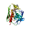
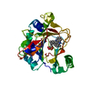
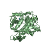
| タイトル | Crystal Structure Analysis of HI1317 |
|---|
 要素 要素 | HI1317 |
|---|
 キーワード キーワード | UNKNOWN FUNCTION / hypothetical protein / Structure 2 Function Project / S2F / Structural Genomics |
|---|
| 機能・相同性 |  機能・相同性情報 機能・相同性情報
glucose-6-phosphate 1-epimerase / glucose-6-phosphate 1-epimerase activity / carbohydrate binding / carbohydrate metabolic process類似検索 - 分子機能 Glucose-6-phosphate 1-epimerase / Aldose 1-/Glucose-6-phosphate 1-epimerase / Aldose 1-epimerase / Beta-galactosidase; Chain A, domain 5 - #10 / Glycoside hydrolase-type carbohydrate-binding / Beta-galactosidase; Chain A, domain 5 / Galactose mutarotase-like domain superfamily / Distorted Sandwich / Mainly Beta類似検索 - ドメイン・相同性 |
|---|
| 生物種 |  Haemophilus influenzae (インフルエンザ菌) Haemophilus influenzae (インフルエンザ菌) |
|---|
| 手法 |  X線回折 / シンクロトロン / 解像度: 1.57 Å X線回折 / シンクロトロン / 解像度: 1.57 Å |
|---|
 データ登録者 データ登録者 | Bonander, N. / Tordova, M. / Howard, A.J. / Eisenstein, E. / Gilliland, G. / Structure 2 Function Project (S2F) |
|---|
 引用 引用 | ジャーナル: TO BE PUBLISHED
タイトル: Crystal 1.57-A Crystal Structure of HI1317
著者: Bonander, N. / Tordova, M. / Howard, A.J. / Eisenstein, E. / Gilliland, G. |
|---|
| 履歴 | | 登録 | 2001年7月31日 | 登録サイト: RCSB / 処理サイト: RCSB |
|---|
| 改定 1.0 | 2003年6月24日 | Provider: repository / タイプ: Initial release |
|---|
| 改定 1.1 | 2008年4月27日 | Group: Version format compliance |
|---|
| 改定 1.2 | 2011年7月13日 | Group: Version format compliance |
|---|
| 改定 1.3 | 2024年11月6日 | Group: Data collection / Database references ...Data collection / Database references / Derived calculations / Structure summary
カテゴリ: chem_comp_atom / chem_comp_bond ...chem_comp_atom / chem_comp_bond / database_2 / pdbx_entry_details / pdbx_modification_feature / struct_site
Item: _database_2.pdbx_DOI / _database_2.pdbx_database_accession ..._database_2.pdbx_DOI / _database_2.pdbx_database_accession / _struct_site.pdbx_auth_asym_id / _struct_site.pdbx_auth_comp_id / _struct_site.pdbx_auth_seq_id |
|---|
|
|---|
 ムービー
ムービー コントローラー


 データを開く
 基本情報
基本情報 構造の表示
構造の表示 ダウンロードとリンク
 その他のダウンロード
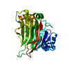


 PDBj
PDBj


 集合体
集合体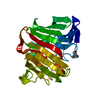

 分子量: 96.063 Da / 分子数: 1 / 由来タイプ: 合成 / 式: SO4
 分子量: 122.143 Da / 分子数: 2 / 由来タイプ: 合成 / 式: C4H12NO3 / コメント: pH緩衝剤*YM
 分子量: 18.015 Da / 分子数: 235 / 由来タイプ: 天然 / 式: H2O
 試料調製
試料調製 / ビームライン: 17-ID / 波長: 1
  解析
解析