 Movie
Movie Controller


+ Open data

- Basic information
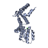
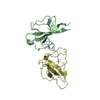
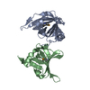
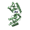
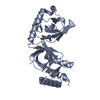
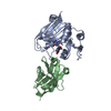
| Entry | Database: PDB / ID: 1jhe | ||||||
|---|---|---|---|---|---|---|---|
| Title | LEXA L89P Q92W E152A K156A MUTANT | ||||||
 Components Components | LEXA REPRESSOR Repressor lexA Repressor lexA | ||||||
 Keywords Keywords | HYDROLASE / LexA SOS repressor / C-terminal | ||||||
| Function / homology |  Function and homology informationrepressor LexA / SOS response / DNA-binding transcription repressor activity / protein-DNA complex / DNA replication / transcription cis-regulatory region binding / serine-type endopeptidase activity / DNA repair / negative regulation of DNA-templated transcription / DNA-templated transcription ...repressor LexA / SOS response / DNA-binding transcription repressor activity / protein-DNA complex / DNA replication / transcription cis-regulatory region binding / serine-type endopeptidase activity / DNA repair / negative regulation of DNA-templated transcription / DNA-templated transcription / DNA damage response / proteolysis / DNA binding / identical protein binding / cytosol Function and homology informationrepressor LexA / SOS response / DNA-binding transcription repressor activity / protein-DNA complex / DNA replication / transcription cis-regulatory region binding / serine-type endopeptidase activity / DNA repair / negative regulation of DNA-templated transcription / DNA-templated transcription ...repressor LexA / SOS response / DNA-binding transcription repressor activity / protein-DNA complex / DNA replication / transcription cis-regulatory region binding / serine-type endopeptidase activity / DNA repair / negative regulation of DNA-templated transcription / DNA-templated transcription / DNA damage response / proteolysis / DNA binding / identical protein binding / cytosolSimilarity search - Function | ||||||
| Biological species |  Escherichia coli (E. coli) Escherichia coli (E. coli) | ||||||
| Method | X-RAY DIFFRACTION / MOLECULAR REPLACEMENT / Resolution: 2.5 Å | ||||||
 Authors Authors | Luo, Y. / Pfuetzner, R.A. / Mosimann, S. / Little, J.W. / J Strynadka, N.C. | ||||||
 Citation Citation | Journal: Cell(Cambridge,Mass.) / Year: 2001 Title: Crystal structure of LexA: a conformational switch for regulation of self-cleavage. Authors: Luo, Y. / Pfuetzner, R.A. / Mosimann, S. / Paetzel, M. / Frey, E.A. / Cherney, M. / Kim, B. / Little, J.W. / Strynadka, N.C. | ||||||
| History |
|
- Structure visualization
| Structure viewer | Molecule: MolmilJmol/JSmol |
|---|
- Downloads & links
-Download
| PDBx/mmCIF format | 1jhe.cif.gz | 59.6 KB | Display | PDBx/mmCIF format |
|---|---|---|---|---|
| PDB format | pdb1jhe.ent.gz | 43.4 KB | Display | PDB format |
| PDBx/mmJSON format | 1jhe.json.gz | Tree view | PDBx/mmJSON format | |
| Others |  Other downloads Other downloads |
-Validation report
| Arichive directory | https://data.pdbj.org/pub/pdb/validation_reports/jh/1jheftp://data.pdbj.org/pub/pdb/validation_reports/jh/1jhe | HTTPS FTP |
|---|
-Related structure data
| Related structure data |  1jhcSC  1jhfC  1jhhC S: Starting model for refinement C: citing same article ( |
|---|---|
| Similar structure data |


-Links
 PDBj
PDBj- Assembly
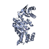
| Deposited unit | 
| ||||||||
|---|---|---|---|---|---|---|---|---|---|
| 1 |
| ||||||||
| Unit cell |
|
-Components
| #1: Protein | Repressor lexA Mass: 14924.055 Da / Num. of mol.: 2 / Fragment: C-Terminus, Residues 68-202 / Mutation: L89P,Q92W,E152A,K156A Source method: isolated from a genetically manipulated source Source: (gene. exp.) Escherichia coli (E. coli) / Gene: LexA / Production host: Escherichia coli (E. coli) / References: UniProt: P0A7C2, repressor LexA#2: Water | ChemComp-HOH / | Water Mass: 18.015 Da / Num. of mol.: 17 / Source method: isolated from a natural source / Formula: H2O Mass: 18.015 Da / Num. of mol.: 17 / Source method: isolated from a natural source / Formula: H2O |
|---|
-Experimental details
-Experiment
| Experiment | Method: X-RAY DIFFRACTION / Number of used crystals: 1 |
|---|
- Sample preparation
| Crystal | Density Matthews: 2.13 Å3/Da / Density % sol: 42.22 % | |||||||||||||||
|---|---|---|---|---|---|---|---|---|---|---|---|---|---|---|---|---|
| Crystal grow | Temperature: 293 K / Method: vapor diffusion, hanging drop / pH: 5.6 Details: 30% PEG 1500, 0.1 M MES buffer, pH 5.6, VAPOR DIFFUSION, HANGING DROP, temperature 293K | |||||||||||||||
| Crystal grow | *PLUS | |||||||||||||||
| Components of the solutions | *PLUS
|
-Data collection
| Diffraction | Mean temperature: 100 K |
|---|---|
| Diffraction source | Source: ROTATING ANODE / Type: RIGAKU RU200 / Wavelength: 1.5418 Å |
| Detector | Type: RIGAKU RAXIS IIC / Detector: IMAGE PLATE / Date: Dec 20, 2000 / Details: mirrors |
| Radiation | Monochromator: GRAPHITE / Protocol: SINGLE WAVELENGTH / Monochromatic (M) / Laue (L): M / Scattering type: x-ray |
| Radiation wavelength | Wavelength: 1.5418 Å / Relative weight: 1 |
| Reflection | Resolution: 2.5→20 Å / Num. all: 9700 / Num. obs: 9700 / % possible obs: 96.9 % / Observed criterion σ(F): 0 / Observed criterion σ(I): 0 / Redundancy: 3.5 % / Rmerge(I) obs: 0.074 / Rsym value: 0.074 / Net I/σ(I): 18.2 |
| Reflection shell | Resolution: 2.5→2.54 Å / Redundancy: 3 % / Rmerge(I) obs: 0.431 / Mean I/σ(I) obs: 2.3 / Num. unique all: 486 / Rsym value: 0.431 / % possible all: 95.1 |
| Reflection | *PLUS Num. obs: 8960 / Num. measured all: 30913 |
| Reflection shell | *PLUS % possible obs: 95.1 % / Num. unique obs: 486 |
- Processing
| Software |
| |||||||||||||||||||||||||
|---|---|---|---|---|---|---|---|---|---|---|---|---|---|---|---|---|---|---|---|---|---|---|---|---|---|---|
| Refinement | Method to determine structure: MOLECULAR REPLACEMENT Starting model: PDB ENTRY 1JHC Resolution: 2.5→15 Å / Isotropic thermal model: Isotropic / Cross valid method: THROUGHOUT / σ(F): 0 / σ(I): 0 / Stereochemistry target values: Engh & Huber
| |||||||||||||||||||||||||
| Refinement step | Cycle: LAST / Resolution: 2.5→15 Å
| |||||||||||||||||||||||||
| Refine LS restraints |
| |||||||||||||||||||||||||
| Software | *PLUS Name: CNS / Version: 1 / Classification: refinement | |||||||||||||||||||||||||
| Refinement | *PLUS Highest resolution: 2.5 Å / Lowest resolution: 15 Å / σ(F): 0 / Rfactor obs: 0.22 / Rfactor Rwork: 0.22 / % reflection Rfree: 10 % | |||||||||||||||||||||||||
| Solvent computation | *PLUS | |||||||||||||||||||||||||
| Displacement parameters | *PLUS |
