 Movie
Movie Controller

[English] 日本語
 Yorodumi
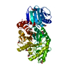
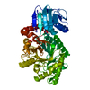
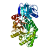
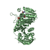
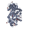
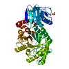
- PDB-1jak: Streptomyces plicatus beta-N-acetylhexosaminidase in Complex with... -
+ Open data

- Basic information
| Entry | Database: PDB / ID: 1jak | |||||||||
|---|---|---|---|---|---|---|---|---|---|---|
| Title | Streptomyces plicatus beta-N-acetylhexosaminidase in Complex with (2R,3R,4S,5R)-2-acetamido-3,4-dihydroxy-5-hydroxymethyl-piperidinium chloride (IFG) | |||||||||
 Components Components | Beta-N-acetylhexosaminidase | |||||||||
 Keywords Keywords | HYDROLASE / glycoside hydrolase / family 20 / Beta-N-acetylhexosaminidase / substrate-assisted catalysis / alpha/beta barrel / isofagomine inhibitor complex | |||||||||
| Function / homology |  Function and homology information Function and homology informationCa2+ activated K+ channels / glycosaminoglycan metabolic process / beta-N-acetylhexosaminidase activity / calcium-activated potassium channel activity / beta-N-acetylhexosaminidase / cGMP effects / ion channel inhibitor activity / action potential / potassium channel regulator activity / detection of calcium ion ...Ca2+ activated K+ channels / glycosaminoglycan metabolic process / beta-N-acetylhexosaminidase activity / calcium-activated potassium channel activity / beta-N-acetylhexosaminidase / cGMP effects / ion channel inhibitor activity / action potential / potassium channel regulator activity / detection of calcium ion / regulation of vasoconstriction / neuronal action potential / voltage-gated potassium channel complex / potassium ion transport / carbohydrate metabolic process / membrane / plasma membrane Similarity search - Function | |||||||||
| Biological species |  Streptomyces plicatus (bacteria) Streptomyces plicatus (bacteria) | |||||||||
| Method |  X-RAY DIFFRACTION / SYNCHROTRON / FOURIER SYNTHESIS / Resolution: 1.75 Å X-RAY DIFFRACTION / SYNCHROTRON / FOURIER SYNTHESIS / Resolution: 1.75 Å | |||||||||
 Authors Authors | Mark, B.L. / Vocadlo, D.J. / Zhao, D. / Knapp, S. / Withers, S.G. / James, M.N. | |||||||||
 Citation Citation | Journal: J.Biol.Chem. / Year: 2001 Title: Biochemical and structural assessment of the 1-N-azasugar GalNAc-isofagomine as a potent family 20 beta-N-acetylhexosaminidase inhibitor. Authors: Mark, B.L. / Vocadlo, D.J. / Zhao, D. / Knapp, S. / Withers, S.G. / James, M.N. #1: Journal: J.Biol.Chem. / Year: 2001Title: Crystallographic Evidence for Substrate-assisted Catalysis in a Bacterial beta-hexosaminidase Authors: Mark, B.L. / Vocadlo, D.J. / Knapp, S. / Triggs-Raine, B.L. / Withers, S.G. / James, M.N. | |||||||||
| History |
|
- Structure visualization
| Structure viewer | Molecule: MolmilJmol/JSmol |
|---|
- Downloads & links
-Download
| PDBx/mmCIF format | 1jak.cif.gz | 124.5 KB | Display | PDBx/mmCIF format |
|---|---|---|---|---|
| PDB format | pdb1jak.ent.gz | 93.2 KB | Display | PDB format |
| PDBx/mmJSON format | 1jak.json.gz | Tree view | PDBx/mmJSON format | |
| Others |  Other downloads Other downloads |
-Validation report
| Arichive directory | https://data.pdbj.org/pub/pdb/validation_reports/ja/1jakftp://data.pdbj.org/pub/pdb/validation_reports/ja/1jak | HTTPS FTP |
|---|
-Related structure data
| Related structure data |  1hp4S S: Starting model for refinement |
|---|---|
| Similar structure data |


-Links
 PDBj
PDBj

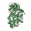
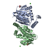
- Assembly
| Deposited unit | 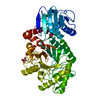
| ||||||||
|---|---|---|---|---|---|---|---|---|---|
| 1 |
| ||||||||
| Unit cell |
| ||||||||
| Components on special symmetry positions |
|
-Components
-Protein , 1 types, 1 molecules A
| #1: Protein | Mass: 56126.777 Da / Num. of mol.: 1 Source method: isolated from a genetically manipulated source Source: (gene. exp.) Streptomyces plicatus (bacteria) / Plasmid: p3AHEX-1.8 / Species (production host): Escherichia coli / Production host: References: UniProt: Q9Y691, UniProt: O85361*PLUS, beta-N-acetylhexosaminidase |
|---|
-Non-polymers , 5 types, 537 molecules 


| #2: Chemical | ChemComp-SO4 /  Mass: 96.063 Da / Num. of mol.: 1 / Source method: obtained synthetically / Formula: SO4 Mass: 96.063 Da / Num. of mol.: 1 / Source method: obtained synthetically / Formula: SO4 | ||||||
|---|---|---|---|---|---|---|---|
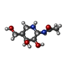
| #3: Chemical |  Mass: 35.453 Da / Num. of mol.: 3 / Source method: obtained synthetically / Formula: Cl Mass: 35.453 Da / Num. of mol.: 3 / Source method: obtained synthetically / Formula: Cl#4: Chemical | ChemComp-IFG / ( |  Mass: 204.224 Da / Num. of mol.: 1 / Source method: obtained synthetically / Formula: C8H16N2O4 Mass: 204.224 Da / Num. of mol.: 1 / Source method: obtained synthetically / Formula: C8H16N2O4#5: Chemical |  Mass: 92.094 Da / Num. of mol.: 3 / Source method: obtained synthetically / Formula: C3H8O3 Mass: 92.094 Da / Num. of mol.: 3 / Source method: obtained synthetically / Formula: C3H8O3#6: Water | ChemComp-HOH / | Mass: 18.015 Da / Num. of mol.: 529 / Source method: isolated from a natural source / Formula: H2O |
-Details
| Has protein modification | Y |
|---|
-Experimental details
-Experiment
| Experiment | Method: X-RAY DIFFRACTION / Number of used crystals: 1 |
|---|
- Sample preparation
| Crystal | Density Matthews: 4.01 Å3/Da / Density % sol: 69.32 % | |||||||||||||||||||||||||||||||||||||||||||||||||
|---|---|---|---|---|---|---|---|---|---|---|---|---|---|---|---|---|---|---|---|---|---|---|---|---|---|---|---|---|---|---|---|---|---|---|---|---|---|---|---|---|---|---|---|---|---|---|---|---|---|---|
| Crystal grow | Temperature: 298 K / Method: vapor diffusion, hanging drop / pH: 6 Details: Ammonium sulphate, Tri-sodium citrate, Sodium Chloride, glycerol, pH 6.0, VAPOR DIFFUSION, HANGING DROP, temperature 298.0K | |||||||||||||||||||||||||||||||||||||||||||||||||
| Crystal grow | *PLUS pH: 5 | |||||||||||||||||||||||||||||||||||||||||||||||||
| Components of the solutions | *PLUS
|
-Data collection
| Diffraction | Mean temperature: 100 K |
|---|---|
| Diffraction source | Source: SYNCHROTRON / Site: SSRL  / Beamline: BL9-1 / Wavelength: 0.979 Å / Beamline: BL9-1 / Wavelength: 0.979 Å |
| Detector | Type: MARRESEARCH / Detector: IMAGE PLATE / Date: Nov 16, 2000 |
| Radiation | Monochromator: single crystal Si(331) bent monochromator / Protocol: SINGLE WAVELENGTH / Monochromatic (M) / Laue (L): M / Scattering type: x-ray |
| Radiation wavelength | Wavelength: 0.979 Å / Relative weight: 1 |
| Reflection | Resolution: 1.75→44.1 Å / Num. all: 92286 / Num. obs: 92238 / % possible obs: 99.6 % / Observed criterion σ(F): 0 / Observed criterion σ(I): -3 / Redundancy: 6 % / Biso Wilson estimate: 12.2 Å2 / Rmerge(I) obs: 0.043 / Net I/σ(I): 36.2 |
| Reflection shell | Resolution: 1.75→1.78 Å / Redundancy: 6 % / Rmerge(I) obs: 0.15 / Mean I/σ(I) obs: 10.9 / Num. unique all: 4366 / % possible all: 95.7 |
| Reflection | *PLUS Lowest resolution: 100 Å / Num. obs: 92286 / Num. measured all: 1013193 |
| Reflection shell | *PLUS % possible obs: 95.7 % / Num. unique obs: 4366 |
- Processing
| Software |
| ||||||||||||||||||||||||||||||||||||||||||||
|---|---|---|---|---|---|---|---|---|---|---|---|---|---|---|---|---|---|---|---|---|---|---|---|---|---|---|---|---|---|---|---|---|---|---|---|---|---|---|---|---|---|---|---|---|---|
| Refinement | Method to determine structure: FOURIER SYNTHESIS Starting model: PDB ENTRY 1HP4 Resolution: 1.75→44.1 Å / Rfactor Rfree error: 0.002 / Data cutoff high absF: 3274409.67 / Data cutoff low absF: 0 / Isotropic thermal model: RESTRAINED / Cross valid method: THROUGHOUT / σ(F): 0 / σ(I): 0 / Stereochemistry target values: Engh & Huber
| ||||||||||||||||||||||||||||||||||||||||||||
| Solvent computation | Solvent model: FLAT MODEL / Bsol: 51.1 Å2 / ksol: 0.391 e/Å3 | ||||||||||||||||||||||||||||||||||||||||||||
| Displacement parameters | Biso mean: 14.4 Å2
| ||||||||||||||||||||||||||||||||||||||||||||
| Refine analyze |
| ||||||||||||||||||||||||||||||||||||||||||||
| Refinement step | Cycle: LAST / Resolution: 1.75→44.1 Å
| ||||||||||||||||||||||||||||||||||||||||||||
| Refine LS restraints |
| ||||||||||||||||||||||||||||||||||||||||||||
| LS refinement shell | Resolution: 1.75→1.86 Å / Rfactor Rfree error: 0.005 / Total num. of bins used: 6
| ||||||||||||||||||||||||||||||||||||||||||||
| Xplor file |
| ||||||||||||||||||||||||||||||||||||||||||||
| Software | *PLUS Name: CNS / Version: 1 / Classification: refinement | ||||||||||||||||||||||||||||||||||||||||||||
| Refinement | *PLUS Lowest resolution: 100 Å / σ(F): 0 / % reflection Rfree: 10.1 % | ||||||||||||||||||||||||||||||||||||||||||||
| Solvent computation | *PLUS | ||||||||||||||||||||||||||||||||||||||||||||
| Displacement parameters | *PLUS Biso mean: 14.4 Å2 | ||||||||||||||||||||||||||||||||||||||||||||
| Refine LS restraints | *PLUS
| ||||||||||||||||||||||||||||||||||||||||||||
| LS refinement shell | *PLUS Rfactor Rfree: 0.204 / % reflection Rfree: 10 % / Rfactor Rwork: 0.184 |
