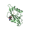
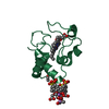
Mass: 2954.955 Da / Num. of mol.: 1 / Source method: obtained synthetically
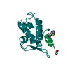
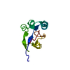
#2: Protein
HeterogeneousnuclearribonucleoproteinK / HNRNP K / DC-stretch binding protein / CSBP / Transformation upregulated nuclear protein / TUNP
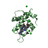
Mass: 9656.836 Da / Num. of mol.: 1 / Fragment: KH3 domain, RESIDUES 379-463, NUMBERED 5-89 Source method: isolated from a genetically manipulated source Source: (gene. exp.) Homo sapiens (human) / Plasmid: PET15B / Production host: Escherichia coli (E. coli) / Strain (production host): BE23 / References: UniProt: P61978
-
Experimental details
-
Experiment
Experiment
Method: SOLUTION NMR Details: (5) IPAP EXPTS FOR DIPOLAR COUPLINGS WERE MEASURED IN A LIQUID CRYSTALLINE MEDIUM OF PHAGE PF1 (18 MG/ML), 5% C12E5 POLYETHYLENE GLYCOL/HEXANOL (MOLAR RATIO OF SURFACTANT TO ALCOHOL 0.96)
(4) 2D 12C-FILTERED EXPERIMENTS FOR DNA ASSIGNMENTS
1
5
1
(5) IPAP EXPTS FOR DIPOLAR COUPLINGS
-
Sample preparation
Sample conditions
Ionic strength: 50 mM SODIUM PHOSPHATE / pH: 6.80 / Temperature: 308 K
Crystal grow
*PLUS
Method: other / Details: NMR
-
NMR measurement
NMR spectrometer
Type
Manufacturer
Model
Field strength (MHz)
Spectrometer-ID
Bruker DMX
Bruker
DMX
600
1
Bruker DRX
Bruker
DRX
750
2
Bruker DRX
Bruker
DRX
800
3
-
Processing
NMR software
Name
Version
Developer
Classification
X-PLOR NIH
(HTTP://NMR.CIT.NIH.GOV/XPLOR_NIH)
CLORE, KUSZEWSKI, SCHWIETERS, TJANDRA
refinement
XPLOR_NIH
structuresolution
Refinement
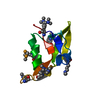
Method: simulated annealing / Software ordinal: 1 Details: THE STRUCTURES WERE CALCULATED BY SIMULATED ANNEALING IN TORSION ANGLE SPACE (SCHWIETERS AND CLORE (2001) J MAGN RESON 152, 288-302) AGAINST A TARGET FUNCTION COMPRISING THE EXPERIMENTAL NMR ...Details: THE STRUCTURES WERE CALCULATED BY SIMULATED ANNEALING IN TORSION ANGLE SPACE (SCHWIETERS AND CLORE (2001) J MAGN RESON 152, 288-302) AGAINST A TARGET FUNCTION COMPRISING THE EXPERIMENTAL NMR RESTRAINTS (NOE-DERIVED INTERPROTON DISTANCE, TORSION ANGLE, 13CALPHA/13CBETA SHIFTS AND DIPOLAR COUPLINGS). THE NON-BONDED CONTACTS IN THE TARGET FUNCTION ARE REPRESENTED BY A QUARTIC VAN DER WAALS REPULSION TERM, SUPPLEMENTED BY TORSION ANGLE (KUSZEWSKI ET AL. J. MAGN. RESON 125, 171-177 (1997)) AND BASE-BASE POSITIONAL (KUSZEWSKI ET AL. J AM CHEM SOC 123, 3903-3918 (2001)) DATABASE POTENTIALS OF MEAN FORCE. IN THIS ENTRY THE LAST NUMERICAL COLUMN IS THE RMS OF THE 125 INDIVIDUAL SIMULATED ANNEALING STRUCTURES ABOUT THE MEAN COORDINATE POSITIONS: RESIDUES 1-9 and 86-89 OF THE PROTEIN ARE DISORDERED IN THE COMPLEX. ALTHOUGH THE SINGLE-STRANDED DNA IS B-LIKE, THE COORDINATES OF THOSE PORTIONS OF THE SS-DNA NOT IN CONTACT WITH THE PROTEIN COULD NOT BE ACCURATELY DETERMINED (BASES 101-104 and 110). THEREFORE ONLY THE COORDINATES OF RESIDUES 10-85 AND NUCLEOTIDES 105-109 ARE PRESENTED. SOLVED BY MULTI HETERONUCLEAR NMR AND IS BASED ON 1986 EXPERIMENTAL NMR RESTRAINTS DISTANCES 1289 TORSION ANGLES 266 13CA/CB SHIFTS 144 1DNH DIPOLARS IN PEG/HEXANOL 63 1DNC' DIPOLARS IN PEG/HEXANOL 44 2DHNC' DIPOLARS IN PEG/HEXANOL 40 1DNH DIPOLARS IN PHAGE PF1 56 1DNC' DIPOLARS IN PHAGE PF1 42 2DHNC' DIPOLARS IN PHAGE PF1 42 BREAKDOWN OF INTRAMOLECULAR PROTEIN DISTANCE RESTRAINTS INTRARESIDUE 315 SEQUENTIAL 282 MEDIUM RANGE 197 LONG RANGE 300 BACKBONE H-BONDS 54 RESTRAINTS FOR 27 H-BONDS INTRA-DNA DISTANCES 68 INTERMOLECULAR DISTANCES 73
NMR ensemble
Conformer selection criteria: REGULARIZED MEAN STRUCTURE / Conformers calculated total number: 125 / Conformers submitted total number: 1
+
About Yorodumi
-
News
-
Feb 9, 2022. New format data for meta-information of EMDB entries
New format data for meta-information of EMDB entries
Version 3 of the EMDB header file is now the official format.
The previous official version 1.9 will be removed from the archive.
In the structure databanks used in Yorodumi, some data are registered as the other names, "COVID-19 virus" and "2019-nCoV". Here are the details of the virus and the list of structure data.
Jan 31, 2019. EMDB accession codes are about to change! (news from PDBe EMDB page)
EMDB accession codes are about to change! (news from PDBe EMDB page)
The allocation of 4 digits for EMDB accession codes will soon come to an end. Whilst these codes will remain in use, new EMDB accession codes will include an additional digit and will expand incrementally as the available range of codes is exhausted. The current 4-digit format prefixed with “EMD-” (i.e. EMD-XXXX) will advance to a 5-digit format (i.e. EMD-XXXXX), and so on. It is currently estimated that the 4-digit codes will be depleted around Spring 2019, at which point the 5-digit format will come into force.
The EM Navigator/Yorodumi systems omit the EMD- prefix.
Related info.:Q: What is EMD? / ID/Accession-code notation in Yorodumi/EM Navigator
Yorodumi is a browser for structure data from EMDB, PDB, SASBDB, etc.
This page is also the successor to EM Navigator detail page, and also detail information page/front-end page for Omokage search.
The word "yorodu" (or yorozu) is an old Japanese word meaning "ten thousand". "mi" (miru) is to see.
Related info.:EMDB / PDB / SASBDB / Comparison of 3 databanks / Yorodumi Search / Aug 31, 2016. New EM Navigator & Yorodumi / Yorodumi Papers / Jmol/JSmol / Function and homology information / Changes in new EM Navigator and Yorodumi
 Movie
Movie Controller

 Yorodumi
 Open data
 Basic information
 Components
 Keywords
 SINGLE-STRANDED DNA BINDING PROTEIN /
  Function and homology information
 Authors
 Citation
 Structure visualization
 Downloads & links
 Other downloads


 PDBj
PDBj


 Assembly

 Sample preparation
 Processing