 Movie
Movie Controller


+ Open data

- Basic information
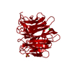
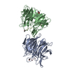
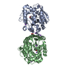
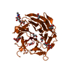
| Entry | Database: PDB / ID: 1iuc | ||||||
|---|---|---|---|---|---|---|---|
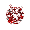
| Title | Fucose-specific lectin from Aleuria aurantia with three ligands | ||||||
 Components Components | Fucose-specific lectin | ||||||
 Keywords Keywords | SUGAR BINDING PROTEIN / Lectin / RIKEN Structural Genomics/Proteomics Initiative / RSGI / Structural Genomics | ||||||
| Function / homology |  Function and homology information Function and homology informationreproductive fruiting body development / fucose binding / defense response to fungus / carbohydrate binding / protein homodimerization activity / identical protein binding Similarity search - Function | ||||||
| Biological species |  Aleuria aurantia (orange peel mushroom) Aleuria aurantia (orange peel mushroom) | ||||||
| Method |  X-RAY DIFFRACTION / SYNCHROTRON / MAD / Resolution: 2.24 Å X-RAY DIFFRACTION / SYNCHROTRON / MAD / Resolution: 2.24 Å | ||||||
 Authors Authors | Fujihashi, M. / Peapus, D.H. / Kamiya, N. / Nagata, Y. / Miki, K. / RIKEN Structural Genomics/Proteomics Initiative (RSGI) | ||||||
 Citation Citation | Journal: Biochemistry / Year: 2003 Title: Crystal Structure of Fucose-Specific Lectin from Aleuria aurantia Binding Ligands at Three of Its Five Sugar Recognition Sites Authors: Fujihashi, M. / Peapus, D.H. / Kamiya, N. / Nagata, Y. / Miki, K. #1: Journal: Acta Crystallogr.,Sect.D / Year: 2003Title: X-ray crystallographic characterization and phasing of a fucose-specific lectin from Aleuria aurantia Authors: Fujihashi, M. / Peapus, D.H. / Nakajima, E. / Yamada, T. / Saito, J.I. / Kita, A. / Higuchi, Y. / Sugawara, Y. / Ando, A. / Kamiya, N. / Nagata, Y. / Miki, K. | ||||||
| History |
|
- Structure visualization
| Structure viewer | Molecule: MolmilJmol/JSmol |
|---|
- Downloads & links
-Download
| PDBx/mmCIF format | 1iuc.cif.gz | 74.8 KB | Display | PDBx/mmCIF format |
|---|---|---|---|---|
| PDB format | pdb1iuc.ent.gz | 56.4 KB | Display | PDB format |
| PDBx/mmJSON format | 1iuc.json.gz | Tree view | PDBx/mmJSON format | |
| Others |  Other downloads Other downloads |
-Validation report
| Arichive directory | https://data.pdbj.org/pub/pdb/validation_reports/iu/1iucftp://data.pdbj.org/pub/pdb/validation_reports/iu/1iuc | HTTPS FTP |
|---|
-Related structure data


-Links
 PDBj
PDBj
- Assembly
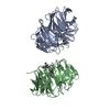
| Deposited unit | 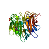
| ||||||||
|---|---|---|---|---|---|---|---|---|---|
| 1 | 
| ||||||||
| Unit cell |
|
-Components
| #1: Protein | Mass: 33425.984 Da / Num. of mol.: 1 Source method: isolated from a genetically manipulated source Source: (gene. exp.) Aleuria aurantia (orange peel mushroom)Plasmid: pKA-1 / Production host:  | ||||||
|---|---|---|---|---|---|---|---|
| #2: Sugar | 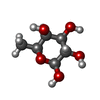  Type: L-saccharide, beta linking / Mass: 164.156 Da / Num. of mol.: 2 Type: L-saccharide, beta linking / Mass: 164.156 Da / Num. of mol.: 2Source method: isolated from a genetically manipulated source Formula: C6H12O5 #3: Sugar | ChemComp-FUC / |   Type: L-saccharide, alpha linking / Mass: 164.156 Da / Num. of mol.: 1 Type: L-saccharide, alpha linking / Mass: 164.156 Da / Num. of mol.: 1Source method: isolated from a genetically manipulated source Formula: C6H12O5 #4: Chemical | ChemComp-SO4 / |   Mass: 96.063 Da / Num. of mol.: 1 / Source method: obtained synthetically / Formula: SO4 Mass: 96.063 Da / Num. of mol.: 1 / Source method: obtained synthetically / Formula: SO4#5: Water | ChemComp-HOH / |  Mass: 18.015 Da / Num. of mol.: 169 / Source method: isolated from a natural source / Formula: H2O Mass: 18.015 Da / Num. of mol.: 169 / Source method: isolated from a natural source / Formula: H2O |
-Experimental details
-Experiment
| Experiment | Method: X-RAY DIFFRACTION / Number of used crystals: 1 |
|---|
- Sample preparation
| Crystal | Density Matthews: 3.81 Å3/Da / Density % sol: 67.72 % | |||||||||||||||||||||||||||||||||||||||||||||||||
|---|---|---|---|---|---|---|---|---|---|---|---|---|---|---|---|---|---|---|---|---|---|---|---|---|---|---|---|---|---|---|---|---|---|---|---|---|---|---|---|---|---|---|---|---|---|---|---|---|---|---|
| Crystal grow | Temperature: 293 K / Method: vapor diffusion, sitting drop / pH: 7.2 Details: potassium phosphate, sodium chloride, potassium chloride, ammonium sulfate, pH 7.2, VAPOR DIFFUSION, SITTING DROP, temperature 293K | |||||||||||||||||||||||||||||||||||||||||||||||||
| Crystal grow | *PLUS Temperature: 297 K / Method: vapor diffusion, sitting dropDetails: Fujihashi, M., (2003) Acta Crystallogr.,Sect.D, 59, 378. | |||||||||||||||||||||||||||||||||||||||||||||||||
| Components of the solutions | *PLUS
|
-Data collection
| Diffraction | Mean temperature: 293 K |
|---|---|
| Diffraction source | Source: SYNCHROTRON / Site: Photon Factory  / Beamline: BL-18B / Wavelength: 1 Å / Beamline: BL-18B / Wavelength: 1 Å |
| Detector | Type: FUJI / Detector: IMAGE PLATE / Date: Jun 16, 1998 |
| Radiation | Monochromator: Silicon (111) / Protocol: MAD / Monochromatic (M) / Laue (L): M / Scattering type: x-ray |
| Radiation wavelength | Wavelength: 1 Å / Relative weight: 1 |
| Reflection | Resolution: 2.24→50 Å / Num. all: 46319 / Num. obs: 46319 / % possible obs: 91.2 % / Observed criterion σ(I): 1 / Redundancy: 8.1 % / Biso Wilson estimate: 22.1 Å2 / Rmerge(I) obs: 0.061 / Net I/σ(I): 31.3 |
| Reflection shell | Resolution: 2.24→2.29 Å / Rmerge(I) obs: 0.301 / % possible all: 81.2 |
| Reflection | *PLUS Lowest resolution: 100 Å |
| Reflection shell | *PLUS % possible obs: 81.2 % |
- Processing
| Software |
| ||||||||||||||||||||||||||||||||||||||||||||||||||||||||||||||||||||||||||||||||
|---|---|---|---|---|---|---|---|---|---|---|---|---|---|---|---|---|---|---|---|---|---|---|---|---|---|---|---|---|---|---|---|---|---|---|---|---|---|---|---|---|---|---|---|---|---|---|---|---|---|---|---|---|---|---|---|---|---|---|---|---|---|---|---|---|---|---|---|---|---|---|---|---|---|---|---|---|---|---|---|---|---|
| Refinement | Method to determine structure: MAD / Resolution: 2.24→39.83 Å / Rfactor Rfree error: 0.004 / Data cutoff high absF: 2387171.8 / Data cutoff low absF: 0 / Isotropic thermal model: RESTRAINED / Cross valid method: THROUGHOUT / σ(F): 0 / Stereochemistry target values: Engh & Huber
| ||||||||||||||||||||||||||||||||||||||||||||||||||||||||||||||||||||||||||||||||
| Solvent computation | Solvent model: FLAT MODEL / Bsol: 46.7864 Å2 / ksol: 0.323438 e/Å3 | ||||||||||||||||||||||||||||||||||||||||||||||||||||||||||||||||||||||||||||||||
| Displacement parameters | Biso mean: 30.6 Å2
| ||||||||||||||||||||||||||||||||||||||||||||||||||||||||||||||||||||||||||||||||
| Refine analyze |
| ||||||||||||||||||||||||||||||||||||||||||||||||||||||||||||||||||||||||||||||||
| Refinement step | Cycle: LAST / Resolution: 2.24→39.83 Å
| ||||||||||||||||||||||||||||||||||||||||||||||||||||||||||||||||||||||||||||||||
| Refine LS restraints |
| ||||||||||||||||||||||||||||||||||||||||||||||||||||||||||||||||||||||||||||||||
| LS refinement shell | Resolution: 2.24→2.38 Å / Rfactor Rfree error: 0.012 / Total num. of bins used: 6
| ||||||||||||||||||||||||||||||||||||||||||||||||||||||||||||||||||||||||||||||||
| Xplor file |
| ||||||||||||||||||||||||||||||||||||||||||||||||||||||||||||||||||||||||||||||||
| Refinement | *PLUS Lowest resolution: 40 Å / Num. reflection obs: 46425 / Rfactor Rwork: 0.165 | ||||||||||||||||||||||||||||||||||||||||||||||||||||||||||||||||||||||||||||||||
| Solvent computation | *PLUS | ||||||||||||||||||||||||||||||||||||||||||||||||||||||||||||||||||||||||||||||||
| Displacement parameters | *PLUS | ||||||||||||||||||||||||||||||||||||||||||||||||||||||||||||||||||||||||||||||||
| Refine LS restraints | *PLUS
| ||||||||||||||||||||||||||||||||||||||||||||||||||||||||||||||||||||||||||||||||
| LS refinement shell | *PLUS Rfactor Rfree: 0.23 / Rfactor Rwork: 0.207 |
