 Movie
Movie Controller

[English] 日本語
 Yorodumi
- PDB-1ofz: Crystal structure of fungal lectin : six-bladed beta-propeller fo... -
+ Open data

- Basic information
| Entry | Database: PDB / ID: 1ofz | ||||||
|---|---|---|---|---|---|---|---|
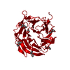
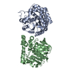
| Title | Crystal structure of fungal lectin : six-bladed beta-propeller fold and novel fucose recognition mode for aleuria aurantia lectin | ||||||
 Components Components | FUCOSE-SPECIFIC LECTIN | ||||||
 Keywords Keywords | SUGAR BINDING PROTEIN / SUGAR-BINDING PROTEIN / LECTIN / AAL / FUCOSE / ALEURIA AURANTIA LECTIN | ||||||
| Function / homology |  Function and homology information Function and homology informationreproductive fruiting body development / fucose binding / defense response to fungus / carbohydrate binding / protein homodimerization activity / identical protein binding Similarity search - Function | ||||||
| Biological species |  ALEURIA AURANTIA (orange peel mushroom) ALEURIA AURANTIA (orange peel mushroom) | ||||||
| Method |  X-RAY DIFFRACTION / SYNCHROTRON / OTHER / Resolution: 1.5 Å X-RAY DIFFRACTION / SYNCHROTRON / OTHER / Resolution: 1.5 Å | ||||||
 Authors Authors | Wimmerova, M. / Mitchell, E. / Sanchez, J.F. / Gautier, C. / Imberty, A. | ||||||
 Citation Citation | Journal: J.Biol.Chem. / Year: 2003 Title: Crystal Structure of Fungal Lectin: Six-Bladed {Beta}-Propeller Fold and Novel Fucose Recognition Mode for Aleuria Aurantia Lectin Authors: Wimmerova, M. / Mitchell, E. / Sanchez, J.F. / Gautier, C. / Imberty, A. | ||||||
| History |
| ||||||
| Remark 700 | SHEET THE SHEET STRUCTURE OF THIS MOLECULE IS BIFURCATED. IN ORDER TO REPRESENT THIS FEATURE IN ... SHEET THE SHEET STRUCTURE OF THIS MOLECULE IS BIFURCATED. IN ORDER TO REPRESENT THIS FEATURE IN THE SHEET RECORDS BELOW, TWO SHEETS ARE DEFINED. |
- Structure visualization
| Structure viewer | Molecule: MolmilJmol/JSmol |
|---|
- Downloads & links
-Download
| PDBx/mmCIF format | 1ofz.cif.gz | 158.1 KB | Display | PDBx/mmCIF format |
|---|---|---|---|---|
| PDB format | pdb1ofz.ent.gz | 127.6 KB | Display | PDB format |
| PDBx/mmJSON format | 1ofz.json.gz | Tree view | PDBx/mmJSON format | |
| Others |  Other downloads Other downloads |
-Validation report
| Arichive directory | https://data.pdbj.org/pub/pdb/validation_reports/of/1ofzftp://data.pdbj.org/pub/pdb/validation_reports/of/1ofz | HTTPS FTP |
|---|
-Related structure data
| Similar structure data |
|---|


-Links
 PDBj
PDBj
- Assembly
| Deposited unit | 
| ||||||||
|---|---|---|---|---|---|---|---|---|---|
| 1 |
| ||||||||
| Unit cell |
| ||||||||
| Noncrystallographic symmetry (NCS) | NCS oper: (Code: given Matrix: (-0.6857, 0.6241, 0.3745), Vector: |
-Components
| #1: Protein | Mass: 33454.039 Da / Num. of mol.: 2 / Source method: isolated from a natural source / Source: (natural) ALEURIA AURANTIA (orange peel mushroom) / References: UniProt: P18891#2: Sugar | ChemComp-FUL /   Type: L-saccharide, beta linking / Mass: 164.156 Da / Num. of mol.: 8 Type: L-saccharide, beta linking / Mass: 164.156 Da / Num. of mol.: 8Source method: isolated from a genetically manipulated source Formula: C6H12O5 #3: Sugar | ChemComp-FUC /   Type: L-saccharide, alpha linking / Mass: 164.156 Da / Num. of mol.: 4 Type: L-saccharide, alpha linking / Mass: 164.156 Da / Num. of mol.: 4Source method: isolated from a genetically manipulated source Formula: C6H12O5 #4: Water | ChemComp-HOH / |  Mass: 18.015 Da / Num. of mol.: 1166 / Source method: isolated from a natural source / Formula: H2O Mass: 18.015 Da / Num. of mol.: 1166 / Source method: isolated from a natural source / Formula: H2OSequence details | P18891 HAS RESIDUE 139 AS GLY AND 163 AS VAL. ELECTRON DENSITY OF MOLECULE CRYSTALLISED INDICATES ...P18891 HAS RESIDUE 139 AS GLY AND 163 AS VAL. ELECTRON DENSITY OF MOLECULE CRYSTALLIS | |
|---|
-Experimental details
-Experiment
| Experiment | Method: X-RAY DIFFRACTION / Number of used crystals: 1 |
|---|
- Sample preparation
| Crystal | Density Matthews: 2.2 Å3/Da / Density % sol: 32.8 % Description: DATA WERE COLLECTED AT ENERGY REMOTE FROM THE MERCURY ANOMALOUS EDGE | ||||||||||||||||||||||||||||||
|---|---|---|---|---|---|---|---|---|---|---|---|---|---|---|---|---|---|---|---|---|---|---|---|---|---|---|---|---|---|---|---|
| Crystal grow | pH: 6.5 Details: 2UL BUFFER (0.1 M SODIUM CACODYLATE PH 6.5, 0.2M MAGNESIUM ACETATE TETRAHYDRATE, 20% PEG 8000) PLUS 2UL OF LECTIN PROTEIN AT 10 MG/ML AND L-FUCOSE AT 137 UG/ML | ||||||||||||||||||||||||||||||
| Crystal | *PLUS Density Matthews: 2.2 Å3/Da / Density % sol: 46 % | ||||||||||||||||||||||||||||||
| Crystal grow | *PLUS Temperature: 20 ℃ / pH: 6.5 / Method: vapor diffusion, hanging drop | ||||||||||||||||||||||||||||||
| Components of the solutions | *PLUS
|
-Data collection
| Diffraction | Mean temperature: 100 K |
|---|---|
| Diffraction source | Source: SYNCHROTRON / Site: ESRF  / Beamline: ID14-1 / Wavelength: 0.934 / Beamline: ID14-1 / Wavelength: 0.934 |
| Detector | Type: ADSC CCD / Detector: CCD / Date: Jan 15, 2003 / Details: MULTILAYER |
| Radiation | Monochromator: DIAMOND / Protocol: SINGLE WAVELENGTH / Monochromatic (M) / Laue (L): M / Scattering type: x-ray |
| Radiation wavelength | Wavelength: 0.934 Å / Relative weight: 1 |
| Reflection | Resolution: 1.5→19.96 Å / Num. obs: 534572 / % possible obs: 96.8 % / Redundancy: 3.6 % / Biso Wilson estimate: 14.4 Å2 / Rmerge(I) obs: 0.049 / Net I/σ(I): 8.4 |
| Reflection shell | Resolution: 1.5→1.55 Å / Redundancy: 2.8 % / Rmerge(I) obs: 0.374 / Mean I/σ(I) obs: 18.4 / % possible all: 96.8 |
| Reflection | *PLUS Highest resolution: 1.5 Å / Num. obs: 94102 / Redundancy: 3.6 % / Num. measured all: 534572 / Rmerge(I) obs: 0.049 |
| Reflection shell | *PLUS Highest resolution: 1.5 Å / % possible obs: 96.8 % / Redundancy: 2.8 % / Rmerge(I) obs: 0.374 / Mean I/σ(I) obs: 1.9 |
- Processing
| Software |
| ||||||||||||||||||||||||||||||||||||||||||||||||||||||||||||||||||||||||||||||||||||||||||||||||||||||||||||||||||||||||||||||||||||||||||||||||||||||||||||||||||||||||||||||||||||||
|---|---|---|---|---|---|---|---|---|---|---|---|---|---|---|---|---|---|---|---|---|---|---|---|---|---|---|---|---|---|---|---|---|---|---|---|---|---|---|---|---|---|---|---|---|---|---|---|---|---|---|---|---|---|---|---|---|---|---|---|---|---|---|---|---|---|---|---|---|---|---|---|---|---|---|---|---|---|---|---|---|---|---|---|---|---|---|---|---|---|---|---|---|---|---|---|---|---|---|---|---|---|---|---|---|---|---|---|---|---|---|---|---|---|---|---|---|---|---|---|---|---|---|---|---|---|---|---|---|---|---|---|---|---|---|---|---|---|---|---|---|---|---|---|---|---|---|---|---|---|---|---|---|---|---|---|---|---|---|---|---|---|---|---|---|---|---|---|---|---|---|---|---|---|---|---|---|---|---|---|---|---|---|---|
| Refinement | Method to determine structure: OTHER / Resolution: 1.5→19.96 Å / Cor.coef. Fo:Fc: 0.977 / Cor.coef. Fo:Fc free: 0.962 / SU B: 1.417 / SU ML: 0.051 / Cross valid method: THROUGHOUT / ESU R: 0.066 / ESU R Free: 0.071 / Stereochemistry target values: MAXIMUM LIKELIHOOD Details: HYDROGENS HAVE BEEN ADDED IN THE RIDING POSITIONS. DISORDERED REGIONS WERE MODELED STEREOCHEMICALLY WITH OCCUPANCY SET TO ZERO.
| ||||||||||||||||||||||||||||||||||||||||||||||||||||||||||||||||||||||||||||||||||||||||||||||||||||||||||||||||||||||||||||||||||||||||||||||||||||||||||||||||||||||||||||||||||||||
| Solvent computation | Ion probe radii: 0.8 Å / Shrinkage radii: 0.8 Å / VDW probe radii: 1.4 Å / Solvent model: BABINET MODEL PLUS MASK | ||||||||||||||||||||||||||||||||||||||||||||||||||||||||||||||||||||||||||||||||||||||||||||||||||||||||||||||||||||||||||||||||||||||||||||||||||||||||||||||||||||||||||||||||||||||
| Displacement parameters | Biso mean: 12.8 Å2
| ||||||||||||||||||||||||||||||||||||||||||||||||||||||||||||||||||||||||||||||||||||||||||||||||||||||||||||||||||||||||||||||||||||||||||||||||||||||||||||||||||||||||||||||||||||||
| Refinement step | Cycle: LAST / Resolution: 1.5→19.96 Å
| ||||||||||||||||||||||||||||||||||||||||||||||||||||||||||||||||||||||||||||||||||||||||||||||||||||||||||||||||||||||||||||||||||||||||||||||||||||||||||||||||||||||||||||||||||||||
| Refine LS restraints |
|