 Movie
Movie Controller


+ Open data

- Basic information
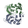
| Entry | Database: PDB / ID: 1isg | |||||||||
|---|---|---|---|---|---|---|---|---|---|---|
| Title | Crystal Structure Analysis of BST-1/CD157 with ATPgammaS | |||||||||
 Components Components | bone marrow stromal cell antigen 1 | |||||||||
 Keywords Keywords | HYDROLASE / ADP ribosylcyclase / NAD glycohydrolase / cns / ATPgammaS | |||||||||
| Function / homology |  Function and homology information Function and homology informationregulation of cellular extravasation / regulation of neutrophil chemotaxis / regulation of superoxide metabolic process / phosphorus-oxygen lyase activity / uropod / Post-translational modification: synthesis of GPI-anchored proteins / regulation of cell-matrix adhesion / Nicotinate metabolism / regulation of integrin-mediated signaling pathway / ADP-ribosyl cyclase/cyclic ADP-ribose hydrolase ...regulation of cellular extravasation / regulation of neutrophil chemotaxis / regulation of superoxide metabolic process / phosphorus-oxygen lyase activity / uropod / Post-translational modification: synthesis of GPI-anchored proteins / regulation of cell-matrix adhesion / Nicotinate metabolism / regulation of integrin-mediated signaling pathway / ADP-ribosyl cyclase/cyclic ADP-ribose hydrolase / NAD+ nucleosidase activity, cyclic ADP-ribose generating / humoral immune response / specific granule membrane / side of membrane / positive regulation of B cell proliferation / regulation of calcium-mediated signaling / regulation of actin cytoskeleton organization / transferase activity / regulation of inflammatory response / positive regulation of cell population proliferation / Neutrophil degranulation / signal transduction / extracellular exosome / extracellular region / plasma membrane Similarity search - Function | |||||||||
| Biological species |  Homo sapiens (human) Homo sapiens (human) | |||||||||
| Method |  X-RAY DIFFRACTION / SYNCHROTRON / MOLECULAR REPLACEMENT / Resolution: 2.6 Å X-RAY DIFFRACTION / SYNCHROTRON / MOLECULAR REPLACEMENT / Resolution: 2.6 Å | |||||||||
 Authors Authors | Yamamoto-Katayama, S. / Ariyoshi, M. / Ishihara, K. / Hirano, T. / Jingami, H. / Morikawa, K. | |||||||||
 Citation Citation | Journal: J.Mol.Biol. / Year: 2002 Title: Crystallographic studies on human BST-1/CD157 with ADP-ribosyl cyclase and NAD glycohydrolase activities. Authors: Yamamoto-Katayama, S. / Ariyoshi, M. / Ishihara, K. / Hirano, T. / Jingami, H. / Morikawa, K. #1: Journal: Biochem.J. / Year: 2001 Title: Site-directed removal of N-glycosylation sites in BST-1/CD157: effects on molecular and functional heterogeneity Authors: Yamamoto-Katayama, S. / Sato, A. / Ariyoshi, M. / Suyama, M. / Ishihara, K. / Hirano, T. / Nakamura, H. / Morikawa, K. / Jingami, H. | |||||||||
| History |
|
- Structure visualization
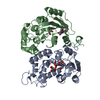
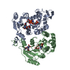
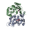
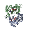
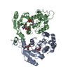
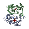
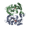
| Structure viewer | Molecule: MolmilJmol/JSmol |
|---|
- Downloads & links
-Download
| PDBx/mmCIF format | 1isg.cif.gz | 117.9 KB | Display | PDBx/mmCIF format |
|---|---|---|---|---|
| PDB format | pdb1isg.ent.gz | 90.5 KB | Display | PDB format |
| PDBx/mmJSON format | 1isg.json.gz | Tree view | PDBx/mmJSON format | |
| Others |  Other downloads Other downloads |
-Validation report
| Arichive directory | https://data.pdbj.org/pub/pdb/validation_reports/is/1isgftp://data.pdbj.org/pub/pdb/validation_reports/is/1isg | HTTPS FTP |
|---|
-Related structure data


-Links
 PDBj
PDBj

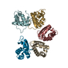
- Assembly
| Deposited unit | 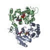
| ||||||||
|---|---|---|---|---|---|---|---|---|---|
| 1 |
| ||||||||
| Unit cell |
|
-Components
| #1: Protein | Mass: 30172.182 Da / Num. of mol.: 2 / Fragment: Extracellular region / Mutation: N34D, N63T, N116A Source method: isolated from a genetically manipulated source Source: (gene. exp.) Homo sapiens (human) / Cell line (production host): High Five / Production host:  Trichoplusia ni (cabbage looper) / References: UniProt: Q10588, NAD+ glycohydrolase Trichoplusia ni (cabbage looper) / References: UniProt: Q10588, NAD+ glycohydrolase#2: Polysaccharide | Source method: isolated from a genetically manipulated source #3: Chemical |   Mass: 523.247 Da / Num. of mol.: 2 / Source method: obtained synthetically / Formula: C10H16N5O12P3S / Comment: ATP-gamma-S, energy-carrying molecule analogue*YM Mass: 523.247 Da / Num. of mol.: 2 / Source method: obtained synthetically / Formula: C10H16N5O12P3S / Comment: ATP-gamma-S, energy-carrying molecule analogue*YM#4: Water | ChemComp-HOH / |  Mass: 18.015 Da / Num. of mol.: 89 / Source method: isolated from a natural source / Formula: H2O Mass: 18.015 Da / Num. of mol.: 89 / Source method: isolated from a natural source / Formula: H2OHas protein modification | Y | |
|---|
-Experimental details
-Experiment
| Experiment | Method: X-RAY DIFFRACTION / Number of used crystals: 1 |
|---|
- Sample preparation
| Crystal | Density Matthews: 3.57 Å3/Da / Density % sol: 65.57 % | |||||||||||||||||||||||||
|---|---|---|---|---|---|---|---|---|---|---|---|---|---|---|---|---|---|---|---|---|---|---|---|---|---|---|
| Crystal grow | Temperature: 293 K / Method: vapor diffusion, hanging drop / pH: 4.5 Details: ammonium sulfate, citrate, ATPgammaS, pH 4.5, VAPOR DIFFUSION, HANGING DROP, temperature 293K | |||||||||||||||||||||||||
| Crystal grow | *PLUS Temperature: 20 ℃ / Method: unknown | |||||||||||||||||||||||||
| Components of the solutions | *PLUS
|
-Data collection
| Diffraction | Mean temperature: 100 K |
|---|---|
| Diffraction source | Source: SYNCHROTRON / Site: SPring-8  / Beamline: BL24XU / Wavelength: 0.836 Å / Beamline: BL24XU / Wavelength: 0.836 Å |
| Detector | Type: RIGAKU RAXIS IV / Detector: IMAGE PLATE / Date: Mar 18, 2000 |
| Radiation | Monochromator: silicon crystal / Protocol: SINGLE WAVELENGTH / Monochromatic (M) / Laue (L): M / Scattering type: x-ray |
| Radiation wavelength | Wavelength: 0.836 Å / Relative weight: 1 |
| Reflection | Resolution: 2.6→20 Å / Num. all: 26538 / Num. obs: 26538 / % possible obs: 98.1 % / Observed criterion σ(F): 0 / Observed criterion σ(I): 0 / Redundancy: 31.2 % / Biso Wilson estimate: 26.9 Å2 / Rmerge(I) obs: 0.068 / Net I/σ(I): 5.9 |
| Reflection shell | Resolution: 2.6→2.69 Å / Rmerge(I) obs: 0.371 / Mean I/σ(I) obs: 33.6 / Num. unique all: 2504 / % possible all: 94.2 |
| Reflection | *PLUS Lowest resolution: 20 Å |
| Reflection shell | *PLUS % possible obs: 94.2 % |
- Processing
| Software |
| ||||||||||||||||||||||||||||||||||||||||||||
|---|---|---|---|---|---|---|---|---|---|---|---|---|---|---|---|---|---|---|---|---|---|---|---|---|---|---|---|---|---|---|---|---|---|---|---|---|---|---|---|---|---|---|---|---|---|
| Refinement | Method to determine structure: MOLECULAR REPLACEMENT / Resolution: 2.6→19.4 Å / Rfactor Rfree error: 0.006 / Isotropic thermal model: RESTRAINED / Cross valid method: THROUGHOUT / σ(F): 0 / σ(I): 0 / Stereochemistry target values: Engh & Huber
| ||||||||||||||||||||||||||||||||||||||||||||
| Solvent computation | Solvent model: FLAT MODEL / Bsol: 34.5738 Å2 / ksol: 0.361196 e/Å3 | ||||||||||||||||||||||||||||||||||||||||||||
| Displacement parameters | Biso mean: 45.1 Å2
| ||||||||||||||||||||||||||||||||||||||||||||
| Refine analyze |
| ||||||||||||||||||||||||||||||||||||||||||||
| Refinement step | Cycle: LAST / Resolution: 2.6→19.4 Å
| ||||||||||||||||||||||||||||||||||||||||||||
| Refine LS restraints |
| ||||||||||||||||||||||||||||||||||||||||||||
| LS refinement shell | Resolution: 2.6→2.76 Å / Rfactor Rfree error: 0.02 / Total num. of bins used: 6
| ||||||||||||||||||||||||||||||||||||||||||||
| Xplor file |
| ||||||||||||||||||||||||||||||||||||||||||||
| Refinement | *PLUS Lowest resolution: 20 Å / σ(F): 0 / % reflection Rfree: 7.5 % / Rfactor obs: 0.211 / Rfactor Rfree: 0.25 | ||||||||||||||||||||||||||||||||||||||||||||
| Solvent computation | *PLUS | ||||||||||||||||||||||||||||||||||||||||||||
| Displacement parameters | *PLUS Biso mean: 45.1 Å2 | ||||||||||||||||||||||||||||||||||||||||||||
| Refine LS restraints | *PLUS
| ||||||||||||||||||||||||||||||||||||||||||||
| LS refinement shell | *PLUS Rfactor Rfree: 0.337 / % reflection Rfree: 7.2 % / Rfactor Rwork: 0.292 |
