 Movie
Movie Controller


+ Open data

- Basic information
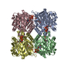
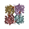
| Entry | Database: PDB / ID: 1iim | ||||||
|---|---|---|---|---|---|---|---|
| Title | thymidylyltransferase complexed with TTP | ||||||
 Components Components | glucose-1-phosphate thymidylyltransferase | ||||||
 Keywords Keywords | TRANSFERASE | ||||||
| Function / homology |  Function and homology information Function and homology informationglucose-1-phosphate thymidylyltransferase / glucose-1-phosphate thymidylyltransferase activity / O antigen biosynthetic process / dTDP-rhamnose biosynthetic process / polysaccharide biosynthetic process / magnesium ion binding / metal ion binding Similarity search - Function | ||||||
| Biological species |  Salmonella enterica (bacteria) Salmonella enterica (bacteria) | ||||||
| Method |  X-RAY DIFFRACTION / Resolution: 2.1 Å X-RAY DIFFRACTION / Resolution: 2.1 Å | ||||||
 Authors Authors | Barton, W.A. / Lesniak, J. / Biggins, J.B. / Jeffrey, P.D. / Jiang, J. / Rajashankar, K.R. / Thorson, J.S. / Nikolov, D.B. | ||||||
 Citation Citation | Journal: Nat.Struct.Biol. / Year: 2001 Title: Structure, mechanism and engineering of a nucleotidylyltransferase as a first step toward glycorandomization. Authors: Barton, W.A. / Lesniak, J. / Biggins, J.B. / Jeffrey, P.D. / Jiang, J. / Rajashankar, K.R. / Thorson, J.S. / Nikolov, D.B. | ||||||
| History |
|
- Structure visualization
| Structure viewer | Molecule: MolmilJmol/JSmol |
|---|
- Downloads & links
-Download
| PDBx/mmCIF format | 1iim.cif.gz | 135.4 KB | Display | PDBx/mmCIF format |
|---|---|---|---|---|
| PDB format | pdb1iim.ent.gz | 106.1 KB | Display | PDB format |
| PDBx/mmJSON format | 1iim.json.gz | Tree view | PDBx/mmJSON format | |
| Others |  Other downloads Other downloads |
-Validation report
| Arichive directory | https://data.pdbj.org/pub/pdb/validation_reports/ii/1iimftp://data.pdbj.org/pub/pdb/validation_reports/ii/1iim | HTTPS FTP |
|---|
-Related structure data


-Links
 PDBj
PDBj


- Assembly
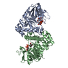
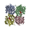
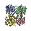
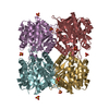
| Deposited unit | 
| ||||||||
|---|---|---|---|---|---|---|---|---|---|
| 1 | 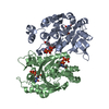
| ||||||||
| Unit cell |
| ||||||||
| Details | The second part of the biological assembly is generated by the two fold axis: -x -y -z+1/2 |
-Components
| #1: Protein | Mass: 32485.201 Da / Num. of mol.: 2 Source method: isolated from a genetically manipulated source Source: (gene. exp.) Salmonella enterica (bacteria) / Strain: LT2 / Plasmid: pET22 / Production host: References: UniProt: Q9F7K6, UniProt: P26393*PLUS, glucose-1-phosphate thymidylyltransferase #2: Chemical | ChemComp-TTP /   Mass: 482.168 Da / Num. of mol.: 4 / Source method: obtained synthetically / Formula: C10H17N2O14P3 Mass: 482.168 Da / Num. of mol.: 4 / Source method: obtained synthetically / Formula: C10H17N2O14P3#3: Water | ChemComp-HOH / |  Mass: 18.015 Da / Num. of mol.: 385 / Source method: isolated from a natural source / Formula: H2O Mass: 18.015 Da / Num. of mol.: 385 / Source method: isolated from a natural source / Formula: H2O |
|---|
-Experimental details
-Experiment
| Experiment | Method: X-RAY DIFFRACTION |
|---|
- Sample preparation
| Crystal | Density Matthews: 2.62 Å3/Da / Density % sol: 53 % | ||||||||||||||||||||||||||||||||||||||||||||||||
|---|---|---|---|---|---|---|---|---|---|---|---|---|---|---|---|---|---|---|---|---|---|---|---|---|---|---|---|---|---|---|---|---|---|---|---|---|---|---|---|---|---|---|---|---|---|---|---|---|---|
| Crystal grow | Temperature: 298 K / Method: vapor diffusion, hanging drop / pH: 8.5 Details: Tris, Mono-ammonium dihydrogen phosphate, pH 8.5, VAPOR DIFFUSION, HANGING DROP, temperature 298K | ||||||||||||||||||||||||||||||||||||||||||||||||
| Crystal grow | *PLUS pH: 7.2 | ||||||||||||||||||||||||||||||||||||||||||||||||
| Components of the solutions | *PLUS
|
-Data collection
| Diffraction | Mean temperature: 120 K |
|---|---|
| Diffraction source | Source: ROTATING ANODE / Type: RIGAKU / Wavelength: 1.5418 |
| Detector | Type: RIGAKU RAXIS IV / Detector: IMAGE PLATE / Details: mirrors |
| Radiation | Protocol: SINGLE WAVELENGTH / Monochromatic (M) / Laue (L): M / Scattering type: x-ray |
| Radiation wavelength | Wavelength: 1.5418 Å / Relative weight: 1 |
| Reflection | Resolution: 2.01→19.26 Å / Num. all: 45400 / Observed criterion σ(F): 0 / Biso Wilson estimate: 18.8 Å2 / Limit h max: 59 / Limit h min: 0 / Limit k max: 40 / Limit k min: 0 / Limit l max: 46 / Limit l min: 0 / Observed criterion F max: 299778.95 / Observed criterion F min: 0.32 |
| Reflection | *PLUS % possible obs: 98.2 % / Redundancy: 9 % / Rmerge(I) obs: 0.109 |
- Processing
| Software |
| ||||||||||||||||||||||||||||||||||||||||||||||||||||||||||||||||||||||||||||||||||||||||||
|---|---|---|---|---|---|---|---|---|---|---|---|---|---|---|---|---|---|---|---|---|---|---|---|---|---|---|---|---|---|---|---|---|---|---|---|---|---|---|---|---|---|---|---|---|---|---|---|---|---|---|---|---|---|---|---|---|---|---|---|---|---|---|---|---|---|---|---|---|---|---|---|---|---|---|---|---|---|---|---|---|---|---|---|---|---|---|---|---|---|---|---|
| Refinement | Resolution: 2.1→19.26 Å / Rfactor Rfree error: 0.003 / Occupancy max: 1 / Occupancy min: 1 / Cross valid method: THROUGHOUT Details: There are three C-terminal residues in the crystal for which the author does not see clear density: LYS 290, GLY 291, LEU 292. There is no density beyond the CB for LYS 154.
| ||||||||||||||||||||||||||||||||||||||||||||||||||||||||||||||||||||||||||||||||||||||||||
| Solvent computation | Solvent model: CNS bulk solvent model used / Bsol: 52.2732 Å2 / ksol: 0.369264 e/Å3 | ||||||||||||||||||||||||||||||||||||||||||||||||||||||||||||||||||||||||||||||||||||||||||
| Displacement parameters | Biso max: 59.32 Å2 / Biso mean: 23.92 Å2 / Biso min: 8.81 Å2
| ||||||||||||||||||||||||||||||||||||||||||||||||||||||||||||||||||||||||||||||||||||||||||
| Refine analyze |
| ||||||||||||||||||||||||||||||||||||||||||||||||||||||||||||||||||||||||||||||||||||||||||
| Refinement step | Cycle: LAST / Resolution: 2.1→19.26 Å
| ||||||||||||||||||||||||||||||||||||||||||||||||||||||||||||||||||||||||||||||||||||||||||
| Refine LS restraints |
| ||||||||||||||||||||||||||||||||||||||||||||||||||||||||||||||||||||||||||||||||||||||||||
| LS refinement shell | Refine-ID: X-RAY DIFFRACTION
| ||||||||||||||||||||||||||||||||||||||||||||||||||||||||||||||||||||||||||||||||||||||||||
| Xplor file |
| ||||||||||||||||||||||||||||||||||||||||||||||||||||||||||||||||||||||||||||||||||||||||||
| Software | *PLUS Name: CNS / Version: 1 / Classification: refinement | ||||||||||||||||||||||||||||||||||||||||||||||||||||||||||||||||||||||||||||||||||||||||||
| Refinement | *PLUS Lowest resolution: 8 Å / Num. reflection obs: 45403 / Num. reflection Rfree: 3583 / % reflection Rfree: 10 % / Rfactor obs: 0.198 | ||||||||||||||||||||||||||||||||||||||||||||||||||||||||||||||||||||||||||||||||||||||||||
| Solvent computation | *PLUS | ||||||||||||||||||||||||||||||||||||||||||||||||||||||||||||||||||||||||||||||||||||||||||
| Displacement parameters | *PLUS | ||||||||||||||||||||||||||||||||||||||||||||||||||||||||||||||||||||||||||||||||||||||||||
| Refine LS restraints | *PLUS
| ||||||||||||||||||||||||||||||||||||||||||||||||||||||||||||||||||||||||||||||||||||||||||
| LS refinement shell | *PLUS Rfactor Rfree: 0.269 / % reflection Rfree: 8.8 % / Rfactor Rwork: 0.268 |
