 Movie
Movie Controller


+ Open data

- Basic information
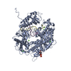
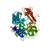
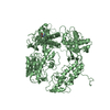
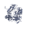
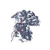
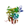
| Entry | Database: PDB / ID: 1ih7 | ||||||
|---|---|---|---|---|---|---|---|
| Title | High-Resolution Structure of Apo RB69 DNA Polymerase | ||||||
 Components Components | DNA POLYMERASE | ||||||
 Keywords Keywords | TRANSFERASE / DNA polymerase / fingers / palm / thumb | ||||||
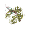
| Function / homology |  Function and homology information Function and homology informationbidirectional double-stranded viral DNA replication / Hydrolases; Acting on ester bonds; Exodeoxyribonucleases producing 5'-phosphomonoesters / 3'-5' exonuclease activity / DNA-templated DNA replication / DNA-directed DNA polymerase / DNA-directed DNA polymerase activity / nucleotide binding / DNA binding / metal ion binding Similarity search - Function | ||||||
| Biological species |  Enterobacteria phage RB69 (virus) Enterobacteria phage RB69 (virus) | ||||||
| Method |  X-RAY DIFFRACTION / SYNCHROTRON / phase extension / Resolution: 2.21 Å X-RAY DIFFRACTION / SYNCHROTRON / phase extension / Resolution: 2.21 Å | ||||||
 Authors Authors | Franklin, M.C. / Wang, J. / Steitz, T.A. | ||||||
 Citation Citation | Journal: Cell(Cambridge,Mass.) / Year: 2001 Title: Structure of the Replicating Complex of a Pol alpha Family DNA Polymerase Authors: Franklin, M.C. / Wang, J. / Steitz, T.A. #1: Journal: Cell(Cambridge,Mass.) / Year: 1997Title: Crystal Structure of a Pol alpha Family Replication DNA Polymerase from Bacteriophage RB69 Authors: Wang, J. / Sattar, A.K. / Wang, C.C. / Karam, J.D. / Konigsberg, W.H. / Steitz, T.A. #2: Journal: Cell(Cambridge,Mass.) / Year: 1999Title: Building a Replisome from Interacting Pieces: Sliding Clamp Complexed to a Peptide from DNA Polymerase and a Polymerase Editing Complex Authors: Shamoo, Y. / Steitz, T.A. | ||||||
| History |
|
- Structure visualization
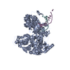
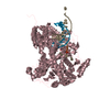
| Structure viewer | Molecule: MolmilJmol/JSmol |
|---|
- Downloads & links
-Download
| PDBx/mmCIF format | 1ih7.cif.gz | 200.8 KB | Display | PDBx/mmCIF format |
|---|---|---|---|---|
| PDB format | pdb1ih7.ent.gz | 157.6 KB | Display | PDB format |
| PDBx/mmJSON format | 1ih7.json.gz | Tree view | PDBx/mmJSON format | |
| Others |  Other downloads Other downloads |
-Validation report
| Arichive directory | https://data.pdbj.org/pub/pdb/validation_reports/ih/1ih7ftp://data.pdbj.org/pub/pdb/validation_reports/ih/1ih7 | HTTPS FTP |
|---|
-Related structure data
| Related structure data | 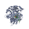 1ig9C  1wajS S: Starting model for refinement C: citing same article ( |
|---|---|
| Similar structure data |

-Links
 PDBj
PDBj


- Assembly
| Deposited unit | 
| ||||||||
|---|---|---|---|---|---|---|---|---|---|
| 1 |
| ||||||||
| Unit cell |
| ||||||||
| Details | Each monomer constitutes a functional assembly. |
-Components
| #1: Protein | Mass: 104743.156 Da / Num. of mol.: 1 Source method: isolated from a genetically manipulated source Source: (gene. exp.) Enterobacteria phage RB69 (virus) / Genus: T4-like viruses / Gene: gp43 / Plasmid: pRB.43 / Species (production host): Escherichia coli / Production host:  | ||||
|---|---|---|---|---|---|
| #2: Chemical |   Mass: 39.098 Da / Num. of mol.: 2 / Source method: obtained synthetically / Formula: K Mass: 39.098 Da / Num. of mol.: 2 / Source method: obtained synthetically / Formula: K#3: Chemical | ChemComp-GMP / | 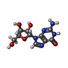  Mass: 283.241 Da / Num. of mol.: 1 / Source method: obtained synthetically / Formula: C10H13N5O5 Mass: 283.241 Da / Num. of mol.: 1 / Source method: obtained synthetically / Formula: C10H13N5O5#4: Water | ChemComp-HOH / |  Mass: 18.015 Da / Num. of mol.: 342 / Source method: isolated from a natural source / Formula: H2O Mass: 18.015 Da / Num. of mol.: 342 / Source method: isolated from a natural source / Formula: H2O |
-Experimental details
-Experiment
| Experiment | Method: X-RAY DIFFRACTION / Number of used crystals: 2 |
|---|
- Sample preparation
| Crystal | Density Matthews: 4.33 Å3/Da / Density % sol: 71.57 % | ||||||||||||||||||||||||||||||||||||
|---|---|---|---|---|---|---|---|---|---|---|---|---|---|---|---|---|---|---|---|---|---|---|---|---|---|---|---|---|---|---|---|---|---|---|---|---|---|
| Crystal grow | Temperature: 285 K / Method: vapor diffusion, hanging drop / pH: 5.6 Details: Sodium citrate, sodium/potassium tartrate, ammonium sulfate, pH 5.6, VAPOR DIFFUSION, HANGING DROP, temperature 285K | ||||||||||||||||||||||||||||||||||||
| Crystal grow | *PLUS Temperature: 16 ℃ / pH: 6.25 / Method: vapor diffusion | ||||||||||||||||||||||||||||||||||||
| Components of the solutions | *PLUS
|
-Data collection
| Diffraction | Mean temperature: 100 K |
|---|---|
| Diffraction source | Source: SYNCHROTRON / Site: CHESS  / Beamline: A1 / Wavelength: 0.913 Å / Beamline: A1 / Wavelength: 0.913 Å |
| Detector | Type: ADSC QUANTUM 4 / Detector: CCD / Date: May 8, 1999 / Details: double crystal monochromator |
| Radiation | Monochromator: double crystal Si(111) / Protocol: SINGLE WAVELENGTH / Monochromatic (M) / Laue (L): M / Scattering type: x-ray |
| Radiation wavelength | Wavelength: 0.913 Å / Relative weight: 1 |
| Reflection | Resolution: 2.2→30 Å / Num. all: 88785 / Num. obs: 88785 / % possible obs: 98.1 % / Observed criterion σ(F): 0 / Observed criterion σ(I): 0 / Redundancy: 5.1 % / Biso Wilson estimate: 31.9 Å2 / Rmerge(I) obs: 0.078 / Net I/σ(I): 21.9 |
| Reflection shell | Resolution: 2.2→2.24 Å / Redundancy: 4.8 % / Rmerge(I) obs: 0.737 / Mean I/σ(I) obs: 3 / Num. unique all: 4352 / % possible all: 98.1 |
| Reflection | *PLUS |
| Reflection shell | *PLUS % possible obs: 98.1 % |
- Processing
| Software |
| ||||||||||||||||||||||||||||||||||||||||
|---|---|---|---|---|---|---|---|---|---|---|---|---|---|---|---|---|---|---|---|---|---|---|---|---|---|---|---|---|---|---|---|---|---|---|---|---|---|---|---|---|---|
| Refinement | Method to determine structure: phase extension Starting model: PDB entry 1WAJ, with all non-protein atoms removed Resolution: 2.21→30 Å / Rfactor Rfree error: 0.003 / Data cutoff high absF: 533579.75 / Data cutoff low absF: 0 / Isotropic thermal model: RESTRAINED / Cross valid method: THROUGHOUT / σ(F): 0 / σ(I): 0 Stereochemistry target values: Engh and Huber, as implemented in CNS Details: This refinement extended the previous apo-RB69 polymerase structure to higher resolution.
| ||||||||||||||||||||||||||||||||||||||||
| Solvent computation | Solvent model: FLAT MODEL / Bsol: 52.24 Å2 / ksol: 0.375 e/Å3 | ||||||||||||||||||||||||||||||||||||||||
| Displacement parameters | Biso mean: 48.9 Å2
| ||||||||||||||||||||||||||||||||||||||||
| Refine analyze |
| ||||||||||||||||||||||||||||||||||||||||
| Refinement step | Cycle: LAST / Resolution: 2.21→30 Å
| ||||||||||||||||||||||||||||||||||||||||
| Refine LS restraints |
| ||||||||||||||||||||||||||||||||||||||||
| LS refinement shell | Resolution: 2.2→2.24 Å / Rfactor Rfree error: 0.027 / Total num. of bins used: 20
| ||||||||||||||||||||||||||||||||||||||||
| Xplor file |
| ||||||||||||||||||||||||||||||||||||||||
| Software | *PLUS Name: CNS / Version: 1 / Classification: refinement | ||||||||||||||||||||||||||||||||||||||||
| Refinement | *PLUS Lowest resolution: 30 Å / σ(F): 0 / % reflection Rfree: 7.9 % / Rfactor all: 10 | ||||||||||||||||||||||||||||||||||||||||
| Solvent computation | *PLUS | ||||||||||||||||||||||||||||||||||||||||
| Displacement parameters | *PLUS Biso mean: 48.9 Å2 | ||||||||||||||||||||||||||||||||||||||||
| Refine LS restraints | *PLUS
| ||||||||||||||||||||||||||||||||||||||||
| LS refinement shell | *PLUS Rfactor Rfree: 0.368 / % reflection Rfree: 10.6 % / Rfactor Rwork: 0.328 |
