 Movie
Movie Controller

[English] 日本語
 Yorodumi
- PDB-1icx: CRYSTAL STRUCTURE OF PATHOGENESIS-RELATED PROTEIN LLPR10.1A FROM ... -
+ Open data

- Basic information
| Entry | Database: PDB / ID: 1icx | ||||||
|---|---|---|---|---|---|---|---|
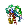
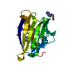
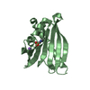
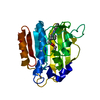
| Title | CRYSTAL STRUCTURE OF PATHOGENESIS-RELATED PROTEIN LLPR10.1A FROM YELLOW LUPINE | ||||||
 Components Components | PROTEIN LLR18A | ||||||
 Keywords Keywords | ALLERGEN / 7-stranded beta sheet / C-terminal helix | ||||||
| Function / homology |  Function and homology information Function and homology informationcytokinin binding / melatonin binding / abscisic acid binding / Hydrolases; Acting on ester bonds; Endoribonucleases producing 3'-phosphomonoesters / abscisic acid-activated signaling pathway / protein phosphatase inhibitor activity / RNA nuclease activity / defense response / signaling receptor activity / hydrolase activity ...cytokinin binding / melatonin binding / abscisic acid binding / Hydrolases; Acting on ester bonds; Endoribonucleases producing 3'-phosphomonoesters / abscisic acid-activated signaling pathway / protein phosphatase inhibitor activity / RNA nuclease activity / defense response / signaling receptor activity / hydrolase activity / calcium ion binding / nucleus / cytosol Similarity search - Function | ||||||
| Biological species |  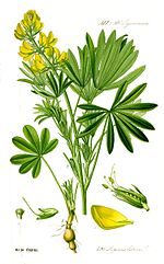 Lupinus luteus (yellow lupine) Lupinus luteus (yellow lupine) | ||||||
| Method |  X-RAY DIFFRACTION / SYNCHROTRON / MOLECULAR REPLACEMENT / Resolution: 1.95 Å X-RAY DIFFRACTION / SYNCHROTRON / MOLECULAR REPLACEMENT / Resolution: 1.95 Å | ||||||
 Authors Authors | Biesiadka, J. / Bujacz, G. / Sikorski, M.M. / Jaskolski, M. | ||||||
 Citation Citation | Journal: J.Mol.Biol. / Year: 2002 Title: Crystal structures of two homologous pathogenesis-related proteins from yellow lupine. Authors: Biesiadka, J. / Bujacz, G. / Sikorski, M.M. / Jaskolski, M. #1: Journal: Acta Crystallogr.,Sect.D / Year: 1999Title: Crystallization and Preliminary X-ray Structure Determination of Lupinus luteus PR10 Protein Authors: Biesiadka, J. / Sikorski, M.M. / Bujacz, G. / Jaskolski, M. #2: Journal: Plant Sci. / Year: 1999Title: Expression of Genes Encoding PR10 Class Pathogenesis-Related Proteins is Inhibited in Yellow Lupine Root Nodules Authors: Sikorski, M.M. / Biesiadka, J. / Kasperska, A.E. / Kopcinska, J. / Lotocka, B. / Golinowski, W. / Legocki, A.B. #3: Journal: Nat.Struct.Biol. / Year: 1996Title: X-ray and NMR Structure of Bet v 1, the Origin of Birch Pollen Allergy Authors: Gajhede, M. / Osmark, P. / Poulsen, F.M. / Ipsen, H. / Larsen, J.N. / Joost van Neerven, R.J. / Schou, C. / Lowenstein, H. / Spangfort, M.D. | ||||||
| History |
|
- Structure visualization
| Structure viewer | Molecule: MolmilJmol/JSmol |
|---|
- Downloads & links
-Download
| PDBx/mmCIF format | 1icx.cif.gz | 43.9 KB | Display | PDBx/mmCIF format |
|---|---|---|---|---|
| PDB format | pdb1icx.ent.gz | 30.3 KB | Display | PDB format |
| PDBx/mmJSON format | 1icx.json.gz | Tree view | PDBx/mmJSON format | |
| Others |  Other downloads Other downloads |
-Validation report
| Arichive directory | https://data.pdbj.org/pub/pdb/validation_reports/ic/1icxftp://data.pdbj.org/pub/pdb/validation_reports/ic/1icx | HTTPS FTP |
|---|
-Related structure data
| Related structure data |  1ifvC  1btvS S: Starting model for refinement C: citing same article ( |
|---|---|
| Similar structure data |


-Links
 PDBj
PDBj- Assembly
| Deposited unit | 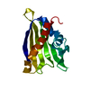
| ||||||||
|---|---|---|---|---|---|---|---|---|---|
| 1 |
| ||||||||
| Unit cell |
|
-Components
| #1: Protein | Mass: 16748.924 Da / Num. of mol.: 1 Source method: isolated from a genetically manipulated source Source: (gene. exp.) Lupinus luteus (yellow lupine) / Plasmid: PET3A-LLPR10.1A / Species (production host): Escherichia coli / Production host:  |
|---|---|
| #2: Water | ChemComp-HOH /  Mass: 18.015 Da / Num. of mol.: 104 / Source method: isolated from a natural source / Formula: H2O Mass: 18.015 Da / Num. of mol.: 104 / Source method: isolated from a natural source / Formula: H2O |
-Experimental details
-Experiment
| Experiment | Method: X-RAY DIFFRACTION / Number of used crystals: 1 |
|---|
- Sample preparation
| Crystal | Density Matthews: 1.83 Å3/Da / Density % sol: 32.8 % |
|---|---|
| Crystal grow | Temperature: 292 K / Method: vapor diffusion, hanging drop / pH: 8 Details: ammonium sulfate, TRIS, sucrose laureate, pH 8.0, VAPOR DIFFUSION, HANGING DROP, temperature 292K |
-Data collection
| Diffraction | Mean temperature: 100 K |
|---|---|
| Diffraction source | Source: SYNCHROTRON / Site: LURE  / Beamline: D41A / Wavelength: 1.375 Å / Beamline: D41A / Wavelength: 1.375 Å |
| Detector | Type: MARRESEARCH / Detector: IMAGE PLATE / Date: Jul 17, 1999 |
| Radiation | Monochromator: Si 111 CHANNEL / Protocol: SINGLE WAVELENGTH / Monochromatic (M) / Laue (L): M / Scattering type: x-ray |
| Radiation wavelength | Wavelength: 1.375 Å / Relative weight: 1 |
| Reflection | Resolution: 1.95→19.35 Å / Num. obs: 9446 / % possible obs: 99.5 % / Observed criterion σ(I): -3 / Redundancy: 9.46 % / Biso Wilson estimate: 12.1 Å2 / Rmerge(I) obs: 0.053 / Net I/σ(I): 25.73 |
| Reflection shell | Resolution: 1.95→2.02 Å / Redundancy: 3.1 % / Rmerge(I) obs: 0.127 / Mean I/σ(I) obs: 8.46 / % possible all: 96.8 |
| Reflection | *PLUS Lowest resolution: 20 Å / Redundancy: 9.5 % / Num. measured all: 89375 / Rmerge(I) obs: 0.053 |
| Reflection shell | *PLUS Lowest resolution: 2.07 Å / % possible obs: 96.8 % / Rmerge(I) obs: 0.127 / Mean I/σ(I) obs: 8.5 |
- Processing
| Software |
| ||||||||||||||||||||||||||||||||||||||||||||||||||||||||||||||||||||||||||||||||
|---|---|---|---|---|---|---|---|---|---|---|---|---|---|---|---|---|---|---|---|---|---|---|---|---|---|---|---|---|---|---|---|---|---|---|---|---|---|---|---|---|---|---|---|---|---|---|---|---|---|---|---|---|---|---|---|---|---|---|---|---|---|---|---|---|---|---|---|---|---|---|---|---|---|---|---|---|---|---|---|---|---|
| Refinement | Method to determine structure: MOLECULAR REPLACEMENT Starting model: PDB ENTRY 1BTV Resolution: 1.95→12 Å / Rfactor Rfree error: 0.007 / Isotropic thermal model: RESTRAINED / Cross valid method: THROUGHOUT / σ(F): 0 / Stereochemistry target values: ENGH & HUBER Details: RESIDUES ASP 60, GLN 133, LYS 135, PHE 136 REFINED AS ALANINES BECAUSE OF POOR SIDE-CHAIN ELECTRON DENSITY
| ||||||||||||||||||||||||||||||||||||||||||||||||||||||||||||||||||||||||||||||||
| Solvent computation | Solvent model: FLAT MODEL / Bsol: 51.41 Å2 / ksol: 0.38 e/Å3 | ||||||||||||||||||||||||||||||||||||||||||||||||||||||||||||||||||||||||||||||||
| Displacement parameters | Biso mean: 26.5 Å2 | ||||||||||||||||||||||||||||||||||||||||||||||||||||||||||||||||||||||||||||||||
| Refine analyze |
| ||||||||||||||||||||||||||||||||||||||||||||||||||||||||||||||||||||||||||||||||
| Refinement step | Cycle: LAST / Resolution: 1.95→12 Å
| ||||||||||||||||||||||||||||||||||||||||||||||||||||||||||||||||||||||||||||||||
| Refine LS restraints |
| ||||||||||||||||||||||||||||||||||||||||||||||||||||||||||||||||||||||||||||||||
| LS refinement shell | Resolution: 1.95→2.07 Å / Rfactor Rfree error: 0.022 / Total num. of bins used: 6
| ||||||||||||||||||||||||||||||||||||||||||||||||||||||||||||||||||||||||||||||||
| Xplor file |
| ||||||||||||||||||||||||||||||||||||||||||||||||||||||||||||||||||||||||||||||||
| Refinement | *PLUS Lowest resolution: 12 Å / % reflection Rfree: 10 % / Rfactor obs: 0.196 / Rfactor Rfree: 0.25 / Rfactor Rwork: 0.196 | ||||||||||||||||||||||||||||||||||||||||||||||||||||||||||||||||||||||||||||||||
| Solvent computation | *PLUS | ||||||||||||||||||||||||||||||||||||||||||||||||||||||||||||||||||||||||||||||||
| Displacement parameters | *PLUS | ||||||||||||||||||||||||||||||||||||||||||||||||||||||||||||||||||||||||||||||||
| Refine LS restraints | *PLUS
| ||||||||||||||||||||||||||||||||||||||||||||||||||||||||||||||||||||||||||||||||
| LS refinement shell | *PLUS Rfactor Rfree: 0.285 / Rfactor Rwork: 0.212 |
