 Movie
Movie Controller

[English] 日本語
 Yorodumi
- PDB-5c9y: Crystal structure of yellow lupine LlPR-10.1A protein partially s... -
+ Open data

- Basic information
| Entry | Database: PDB / ID: 5c9y | ||||||
|---|---|---|---|---|---|---|---|
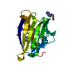
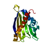
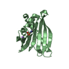
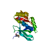
| Title | Crystal structure of yellow lupine LlPR-10.1A protein partially saturated with trans-zeatin | ||||||
 Components Components | Protein LlR18A | ||||||
 Keywords Keywords | PLANT PROTEIN / PR-10 FOLD / LIGAND BINDING / PHYTOHORMONE BINDING PROTEIN / TRANS-ZEATIN / CYTOKININ | ||||||
| Function / homology |  Function and homology information Function and homology informationcytokinin binding / melatonin binding / abscisic acid binding / Hydrolases; Acting on ester bonds; Endoribonucleases producing 3'-phosphomonoesters / abscisic acid-activated signaling pathway / protein phosphatase inhibitor activity / RNA nuclease activity / defense response / signaling receptor activity / hydrolase activity ...cytokinin binding / melatonin binding / abscisic acid binding / Hydrolases; Acting on ester bonds; Endoribonucleases producing 3'-phosphomonoesters / abscisic acid-activated signaling pathway / protein phosphatase inhibitor activity / RNA nuclease activity / defense response / signaling receptor activity / hydrolase activity / calcium ion binding / nucleus / cytosol Similarity search - Function | ||||||
| Biological species |  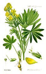 Lupinus luteus (yellow lupine) Lupinus luteus (yellow lupine) | ||||||
| Method |  X-RAY DIFFRACTION / SYNCHROTRON / MOLECULAR REPLACEMENT / Resolution: 1.5 Å X-RAY DIFFRACTION / SYNCHROTRON / MOLECULAR REPLACEMENT / Resolution: 1.5 Å | ||||||
 Authors Authors | Sliwiak, J. / Sikorski, M.M. / Jaskolski, M. | ||||||
 Citation Citation | Journal: J.Struct.Biol. / Year: 2016 Title: Crystallographic and CD probing of ligand-induced conformational changes in a plant PR-10 protein. Authors: Sliwiak, J. / Dolot, R. / Michalska, K. / Szpotkowski, K. / Bujacz, G. / Sikorski, M. / Jaskolski, M. | ||||||
| History |
|
- Structure visualization
| Structure viewer | Molecule: MolmilJmol/JSmol |
|---|
- Downloads & links
-Download
| PDBx/mmCIF format | 5c9y.cif.gz | 78.9 KB | Display | PDBx/mmCIF format |
|---|---|---|---|---|
| PDB format | pdb5c9y.ent.gz | 59.2 KB | Display | PDB format |
| PDBx/mmJSON format | 5c9y.json.gz | Tree view | PDBx/mmJSON format | |
| Others |  Other downloads Other downloads |
-Validation report
| Arichive directory | https://data.pdbj.org/pub/pdb/validation_reports/c9/5c9yftp://data.pdbj.org/pub/pdb/validation_reports/c9/5c9y | HTTPS FTP |
|---|
-Related structure data
| Related structure data |  4ryvC  4y31C  1icxS S: Starting model for refinement C: citing same article ( |
|---|---|
| Similar structure data |


-Links
 PDBj
PDBj- Assembly
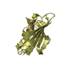
| Deposited unit | 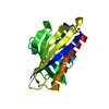
| ||||||||
|---|---|---|---|---|---|---|---|---|---|
| 1 |
| ||||||||
| Unit cell |
|
-Components
| #1: Protein | Mass: 16748.924 Da / Num. of mol.: 1 Source method: isolated from a genetically manipulated source Source: (gene. exp.) Lupinus luteus (yellow lupine) / Gene: LLR18A / Plasmid: PET-3a / Production host:  |
|---|---|
| #2: Water | ChemComp-HOH /  Mass: 18.015 Da / Num. of mol.: 144 / Source method: isolated from a natural source / Formula: H2O Mass: 18.015 Da / Num. of mol.: 144 / Source method: isolated from a natural source / Formula: H2O |
-Experimental details
-Experiment
| Experiment | Method: X-RAY DIFFRACTION |
|---|
- Sample preparation
| Crystal | Density Matthews: 1.92 Å3/Da / Density % sol: 35.86 % / Description: plate |
|---|---|
| Crystal grow | Temperature: 292 K / Method: vapor diffusion, hanging drop / pH: 6.5 / Details: 1.8 M (NH4)2SO4, 0.1 M MES, PH 6.5 |
-Data collection
| Diffraction | Mean temperature: 100 K |
|---|---|
| Diffraction source | Source: SYNCHROTRON / Site: EMBL/DESY, HAMBURG  / Beamline: X11 / Wavelength: 0.81738 Å / Beamline: X11 / Wavelength: 0.81738 Å |
| Detector | Type: MAR555 FLAT PANEL / Detector: IMAGE PLATE / Date: Jul 4, 2011 |
| Radiation | Protocol: SINGLE WAVELENGTH / Monochromatic (M) / Laue (L): M / Scattering type: x-ray |
| Radiation wavelength | Wavelength: 0.81738 Å / Relative weight: 1 |
| Reflection | Resolution: 1.5→50 Å / Num. obs: 21584 / % possible obs: 99.5 % / Redundancy: 7.16 % / Rmerge(I) obs: 0.09 / Net I/σ(I): 21.96 |
| Reflection shell | Resolution: 1.5→1.59 Å / Redundancy: 7.16 % / Rmerge(I) obs: 0.829 / Mean I/σ(I) obs: 2.45 / % possible all: 98.2 |
- Processing
| Software |
| ||||||||||||||||
|---|---|---|---|---|---|---|---|---|---|---|---|---|---|---|---|---|---|
| Refinement | Method to determine structure: MOLECULAR REPLACEMENT Starting model: 1ICX Resolution: 1.5→42.48 Å / Cross valid method: FREE R-VALUE Details: Flat postive electron densities could be observed in the structure indicating the presence of puring rings and tails of three trans-zeatin molecules: two inside protein cavity (one ...Details: Flat postive electron densities could be observed in the structure indicating the presence of puring rings and tails of three trans-zeatin molecules: two inside protein cavity (one interacting with Tyr82 and other with Ser101) and one on the surface of protein (staying in contact with Glu35). However they possess discontinuities and are insufficient for proper ligand modeling thus the structure is deposited without ligand molecules in its coordinates although trans-zeatin was present in crystallization condintions.
| ||||||||||||||||
| Refinement step | Cycle: LAST / Resolution: 1.5→42.48 Å
| ||||||||||||||||
| LS refinement shell | Highest resolution: 1.5 Å |
