 Movie
Movie Controller

[English] 日本語
 Yorodumi
- PDB-1i6q: Formation of a protein intermediate and its trapping by the simul... -
+ Open data

- Basic information
| Entry | Database: PDB / ID: 1i6q | ||||||
|---|---|---|---|---|---|---|---|
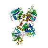
| Title | Formation of a protein intermediate and its trapping by the simultaneous crystallization process: Crystal structure of an iron-saturated intermediate in the FE3+ binding pathway of camel lactoferrin at 2.7 resolution | ||||||
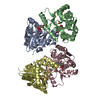
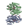
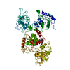
 Components Components | LACTOFERRIN | ||||||
 Keywords Keywords | METAL TRANSPORT / camel lactoferrin / crystal structure / X-ray diffraction / transferrin / lactoferrin / intermediate | ||||||
| Function / homology |  Function and homology information Function and homology informationnegative regulation of tumor necrosis factor (ligand) superfamily member 11 production / negative regulation of single-species biofilm formation in or on host organism / positive regulation of bone mineralization involved in bone maturation / negative regulation of lipopolysaccharide-mediated signaling pathway / specific granule / negative regulation of osteoclast development / antifungal humoral response / positive regulation of chondrocyte proliferation / regulation of tumor necrosis factor production / bone morphogenesis ...negative regulation of tumor necrosis factor (ligand) superfamily member 11 production / negative regulation of single-species biofilm formation in or on host organism / positive regulation of bone mineralization involved in bone maturation / negative regulation of lipopolysaccharide-mediated signaling pathway / specific granule / negative regulation of osteoclast development / antifungal humoral response / positive regulation of chondrocyte proliferation / regulation of tumor necrosis factor production / bone morphogenesis / positive regulation of osteoblast proliferation / Hydrolases; Acting on peptide bonds (peptidases); Serine endopeptidases / positive regulation of osteoblast differentiation / regulation of cytokine production / ossification / innate immune response in mucosa / antibacterial humoral response / iron ion transport / iron ion binding / serine-type endopeptidase activity / negative regulation of apoptotic process / proteolysis / extracellular spaceSimilarity search - Function | ||||||
| Biological species |  Camelus dromedarius (Arabian camel) Camelus dromedarius (Arabian camel) | ||||||
| Method | X-RAY DIFFRACTION / MOLECULAR REPLACEMENT / Resolution: 2.7 Å | ||||||
 Authors Authors | Khan, J.A. / Kumar, P. / Srinivasan, A. / Singh, T.P. | ||||||
 Citation Citation | Journal: J.Biol.Chem. / Year: 2001 Title: Protein intermediate trapped by the simultaneous crystallization process. Crystal structure of an iron-saturated intermediate in the Fe3+ binding pathway of camel lactoferrin at 2.7 a resolution. Authors: Khan, J.A. / Kumar, P. / Srinivasan, A. / Singh, T.P. | ||||||
| History |
| ||||||
| Remark 999 | SEQUENCE THE RESIDUES IN SEQADV HAD BEEN MODELLED BASED ON THE ELECTRON DENSITY MAPS FOLLOWING THE ...SEQUENCE THE RESIDUES IN SEQADV HAD BEEN MODELLED BASED ON THE ELECTRON DENSITY MAPS FOLLOWING THE REPEATED CYCLES OF REFINEMENT |
- Structure visualization
| Structure viewer | Molecule: MolmilJmol/JSmol |
|---|
- Downloads & links
-Download
| PDBx/mmCIF format | 1i6q.cif.gz | 155.7 KB | Display | PDBx/mmCIF format |
|---|---|---|---|---|
| PDB format | pdb1i6q.ent.gz | 117.7 KB | Display | PDB format |
| PDBx/mmJSON format | 1i6q.json.gz | Tree view | PDBx/mmJSON format | |
| Others |  Other downloads Other downloads |
-Validation report
| Arichive directory | https://data.pdbj.org/pub/pdb/validation_reports/i6/1i6qftp://data.pdbj.org/pub/pdb/validation_reports/i6/1i6q | HTTPS FTP |
|---|
-Related structure data
| Related structure data |  1dtzS S: Starting model for refinement |
|---|---|
| Similar structure data |


-Links
 PDBj
PDBj


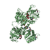
- Assembly
| Deposited unit | 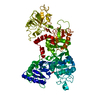
| ||||||||
|---|---|---|---|---|---|---|---|---|---|
| 1 |
| ||||||||
| Unit cell |
|
-Components
| #1: Protein | Mass: 75452.148 Da / Num. of mol.: 1 / Source method: isolated from a natural source / Details: COLOSTRUM / Source: (natural) Camelus dromedarius (Arabian camel) / References: UniProt: Q9TUM0 | ||||
|---|---|---|---|---|---|
| #2: Chemical | Iron  Mass: 55.845 Da / Num. of mol.: 2 / Source method: obtained synthetically / Formula: Fe Mass: 55.845 Da / Num. of mol.: 2 / Source method: obtained synthetically / Formula: Fe#3: Chemical | Carbonate  Mass: 60.009 Da / Num. of mol.: 2 / Source method: obtained synthetically / Formula: CO3 Mass: 60.009 Da / Num. of mol.: 2 / Source method: obtained synthetically / Formula: CO3#4: Water | ChemComp-HOH / | Water Mass: 18.015 Da / Num. of mol.: 233 / Source method: isolated from a natural source / Formula: H2O Mass: 18.015 Da / Num. of mol.: 233 / Source method: isolated from a natural source / Formula: H2O |
-Experimental details
-Experiment
| Experiment | Method: X-RAY DIFFRACTION / Number of used crystals: 1 |
|---|
- Sample preparation
| Crystal | Density Matthews: 2.64 Å3/Da / Density % sol: 53.46 % | |||||||||||||||||||||||||
|---|---|---|---|---|---|---|---|---|---|---|---|---|---|---|---|---|---|---|---|---|---|---|---|---|---|---|
| Crystal grow | Temperature: 277 K / Method: microdialysis / pH: 8 / Details: ethanol, pH 8.0, MICRODIALYSIS, temperature 277K | |||||||||||||||||||||||||
| Crystal grow | *PLUS Temperature: 4 ℃ | |||||||||||||||||||||||||
| Components of the solutions | *PLUS
|
-Data collection
| Diffraction | Mean temperature: 288 K |
|---|---|
| Diffraction source | Source: ROTATING ANODE / Type: RIGAKU RU200 / Wavelength: 1.5418 Å |
| Detector | Type: MARRESEARCH / Detector: IMAGE PLATE / Date: Jul 7, 2000 |
| Radiation | Monochromator: graphite / Protocol: SINGLE WAVELENGTH / Monochromatic (M) / Laue (L): M / Scattering type: x-ray |
| Radiation wavelength | Wavelength: 1.5418 Å / Relative weight: 1 |
| Reflection | Resolution: 2.7→25 Å / Num. all: 44276 / Num. obs: 19901 / % possible obs: 92 % / Observed criterion σ(F): 0 / Observed criterion σ(I): 0 / Redundancy: 2.2 % / Biso Wilson estimate: 35.8 Å2 / Rmerge(I) obs: 0.105 / Rsym value: 0.105 / Net I/σ(I): 9.6 |
| Reflection shell | Resolution: 2.7→2.8 Å / Redundancy: 2.2 % / Rmerge(I) obs: 0.25 / Mean I/σ(I) obs: 6.1 / Num. unique all: 19901 / Rsym value: 0.25 / % possible all: 47 |
| Reflection | *PLUS Num. measured all: 44276 |
| Reflection shell | *PLUS % possible obs: 47 % / Rmerge(I) obs: 0.25 / Mean I/σ(I) obs: 5.6 |
- Processing
| Software |
| |||||||||||||||||||||||||
|---|---|---|---|---|---|---|---|---|---|---|---|---|---|---|---|---|---|---|---|---|---|---|---|---|---|---|
| Refinement | Method to determine structure: MOLECULAR REPLACEMENT Starting model: PDB ENTRY 1DTZ Resolution: 2.7→25 Å / Rfactor Rfree error: 0.008 / Data cutoff high absF: 454981.13 / Data cutoff low absF: 0 / Isotropic thermal model: RESTRAINED / Cross valid method: THROUGHOUT / σ(F): 0 / σ(I): 0.01
| |||||||||||||||||||||||||
| Solvent computation | Solvent model: FLAT MODEL / Bsol: 300 Å2 / ksol: 0.397 e/Å3 | |||||||||||||||||||||||||
| Displacement parameters | Biso mean: 57.1 Å2
| |||||||||||||||||||||||||
| Refine analyze |
| |||||||||||||||||||||||||
| Refinement step | Cycle: LAST / Resolution: 2.7→25 Å
| |||||||||||||||||||||||||
| Refine LS restraints |
| |||||||||||||||||||||||||
| LS refinement shell | Resolution: 2.7→2.87 Å / Rfactor Rfree error: 0.025 / Total num. of bins used: 6
| |||||||||||||||||||||||||
| Xplor file |
| |||||||||||||||||||||||||
| Software | *PLUS Name: CNS / Version: 0.9 / Classification: refinement | |||||||||||||||||||||||||
| Refinement | *PLUS Highest resolution: 2.7 Å / Lowest resolution: 25 Å / σ(F): 0 / % reflection Rfree: 4.8 % | |||||||||||||||||||||||||
| Solvent computation | *PLUS | |||||||||||||||||||||||||
| Displacement parameters | *PLUS Biso mean: 57.1 Å2 | |||||||||||||||||||||||||
| Refine LS restraints | *PLUS
| |||||||||||||||||||||||||
| LS refinement shell | *PLUS Rfactor Rfree: 0.3 / % reflection Rfree: 5.1 % / Rfactor Rwork: 0.28 |
