 Movie
Movie Controller

[English] 日本語
 Yorodumi
- PDB-1hbg: GLYCERA DIBRANCHIATA HEMOGLOBIN. STRUCTURE AND REFINEMENT AT 1.5 ... -
+ Open data

- Basic information
| Entry | Database: PDB / ID: 1hbg | ||||||
|---|---|---|---|---|---|---|---|
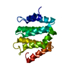
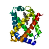
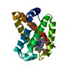
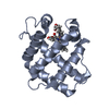
| Title | GLYCERA DIBRANCHIATA HEMOGLOBIN. STRUCTURE AND REFINEMENT AT 1.5 ANGSTROMS RESOLUTION | ||||||
 Components Components | HEMOGLOBIN (CARBONMONOXY) | ||||||
 Keywords Keywords |  OXYGEN TRANSPORT OXYGEN TRANSPORT | ||||||
| Function / homology |  Function and homology informationoxygen carrier activity / oxygen binding / heme binding / metal ion binding Function and homology informationoxygen carrier activity / oxygen binding / heme binding / metal ion bindingSimilarity search - Function | ||||||
| Biological species |  Glycera dibranchiata (invertebrata) Glycera dibranchiata (invertebrata) | ||||||
| Method | X-RAY DIFFRACTION / Resolution: 1.5 Å | ||||||
 Authors Authors | Arents, G.A. / Braden, B.C. / Padlan, E.A. / Love, W.E. | ||||||
 Citation Citation | Journal: J.Mol.Biol. / Year: 1989 Title: Glycera dibranchiata hemoglobin. Structure and refinement at 1.5 A resolution. Authors: Arents, G. / Love, W.E. | ||||||
| History |
|
- Structure visualization
| Structure viewer | Molecule: MolmilJmol/JSmol |
|---|
- Downloads & links
-Download
| PDBx/mmCIF format | 1hbg.cif.gz | 45.3 KB | Display | PDBx/mmCIF format |
|---|---|---|---|---|
| PDB format | pdb1hbg.ent.gz | 31 KB | Display | PDB format |
| PDBx/mmJSON format | 1hbg.json.gz | Tree view | PDBx/mmJSON format | |
| Others |  Other downloads Other downloads |
-Validation report
| Arichive directory | https://data.pdbj.org/pub/pdb/validation_reports/hb/1hbgftp://data.pdbj.org/pub/pdb/validation_reports/hb/1hbg | HTTPS FTP |
|---|
-Related structure data


-Links
 PDBj
PDBj


- Assembly
| Deposited unit | 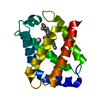
| ||||||||
|---|---|---|---|---|---|---|---|---|---|
| 1 |
| ||||||||
| Unit cell |
| ||||||||
| Atom site foot note | 1: SEVEN RESIDUES HAVE THEIR SIDE CHAINS MODELED IN MORE THAN ONE CONFORMATION. THEY ARE LEU 31, MET 79, LYS 84, ASN 95, SER 109, SER 112, AND SER 143. ALSO SEE REMARK 6 ABOVE. |
-Components
| #1: Protein | Mass: 14935.230 Da / Num. of mol.: 1 Source method: isolated from a genetically manipulated source Source: (gene. exp.) Glycera dibranchiata (invertebrata) / References: UniProt: P02216 |
|---|---|
| #2: Chemical | ChemComp-HEM / Heme B  Mass: 616.487 Da / Num. of mol.: 1 / Source method: obtained synthetically / Formula: C34H32FeN4O4 Mass: 616.487 Da / Num. of mol.: 1 / Source method: obtained synthetically / Formula: C34H32FeN4O4 |
| #3: Chemical | ChemComp-CMO / Carbon monoxide  Mass: 28.010 Da / Num. of mol.: 1 / Source method: obtained synthetically / Formula: CO Mass: 28.010 Da / Num. of mol.: 1 / Source method: obtained synthetically / Formula: CO |
| #4: Water | ChemComp-HOH / Water Mass: 18.015 Da / Num. of mol.: 155 / Source method: isolated from a natural source / Formula: H2O Mass: 18.015 Da / Num. of mol.: 155 / Source method: isolated from a natural source / Formula: H2O |
| Sequence details | THERE ARE A NUMBER OF SEQUENCE DIFFERENCES BETWEEN THE SEQUENCE PRESENTED IN THIS ENTRY AND THAT IN ...THERE ARE A NUMBER OF SEQUENCE DIFFERENCE |
-Experimental details
-Experiment
| Experiment | Method: X-RAY DIFFRACTION |
|---|
- Sample preparation
| Crystal | Density Matthews: 2.3 Å3/Da / Density % sol: 46.52 % | ||||||||||||||||||||||||
|---|---|---|---|---|---|---|---|---|---|---|---|---|---|---|---|---|---|---|---|---|---|---|---|---|---|
| Crystal grow | *PLUS Temperature: 4 ℃ / pH: 6.8 / Method: unknown / Details: took Padlan & Love (1974) from original paper | ||||||||||||||||||||||||
| Components of the solutions | *PLUS
|
-Data collection
| Radiation | Scattering type: x-ray |
|---|---|
| Radiation wavelength | Relative weight: 1 |
| Reflection | *PLUS Highest resolution: 1.5 Å / Lowest resolution: 5 Å / Num. obs: 15951 / Observed criterion σ(I): 1.5 / Num. measured all: 39770 |
- Processing
| Software | Name: PROLSQ / Classification: refinement | |||||||||||||||||||||||||||||||||||||||||||||||||||||||||||||||
|---|---|---|---|---|---|---|---|---|---|---|---|---|---|---|---|---|---|---|---|---|---|---|---|---|---|---|---|---|---|---|---|---|---|---|---|---|---|---|---|---|---|---|---|---|---|---|---|---|---|---|---|---|---|---|---|---|---|---|---|---|---|---|---|---|
| Refinement | Rfactor obs: 0.146 / Highest resolution: 1.5 Å Details: LEUCINE 31 SHOWS EQUAL WEIGHT FOR EACH OF THE THREE POSSIBLE GAUCHE CONFORMATIONS OF ATOMS CD1 AND CD2. THESE ARE PRESENTED AS THREE ALTERNATE CONFORMATIONS WITH AN OCCUPANCY OF 0.33 EACH. ...Details: LEUCINE 31 SHOWS EQUAL WEIGHT FOR EACH OF THE THREE POSSIBLE GAUCHE CONFORMATIONS OF ATOMS CD1 AND CD2. THESE ARE PRESENTED AS THREE ALTERNATE CONFORMATIONS WITH AN OCCUPANCY OF 0.33 EACH. NOTE THAT ONLY THREE SETS OF COORDINATES WERE USED TO MODEL THIS DISORDER AND, THEREFORE, THE SIX ATOMS APPEAR AS THREE ATOMS IN A GRAPHICAL VIEW OF THE ENTRY. | |||||||||||||||||||||||||||||||||||||||||||||||||||||||||||||||
| Refinement step | Cycle: LAST / Highest resolution: 1.5 Å
| |||||||||||||||||||||||||||||||||||||||||||||||||||||||||||||||
| Refine LS restraints |
| |||||||||||||||||||||||||||||||||||||||||||||||||||||||||||||||
| Software | *PLUS Name: PROLSQ / Classification: refinement | |||||||||||||||||||||||||||||||||||||||||||||||||||||||||||||||
| Refinement | *PLUS Highest resolution: 1.5 Å / Rfactor obs: 0.146 | |||||||||||||||||||||||||||||||||||||||||||||||||||||||||||||||
| Solvent computation | *PLUS | |||||||||||||||||||||||||||||||||||||||||||||||||||||||||||||||
| Displacement parameters | *PLUS |
