 Movie
Movie Controller

[English] 日本語
 Yorodumi
- PDB-1jf4: Crystal Structure Of Component IV Glycera Dibranchiata Monomeric ... -
+ Open data

- Basic information
| Entry | Database: PDB / ID: 1jf4 | ||||||
|---|---|---|---|---|---|---|---|
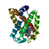
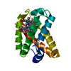
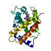
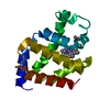
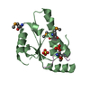
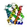
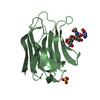
| Title | Crystal Structure Of Component IV Glycera Dibranchiata Monomeric Hemoglobin | ||||||
 Components Components | monomer hemoglobin component IV | ||||||
 Keywords Keywords | OXYGEN STORAGE/TRANSPORT / Glycera / monomer hemoglobin / OXYGEN STORAGE-TRANSPORT COMPLEX | ||||||
| Function / homology |  Function and homology information Function and homology informationoxygen carrier activity / oxygen binding / heme binding / metal ion binding Similarity search - Function | ||||||
| Biological species |  Glycera dibranchiata (invertebrata) Glycera dibranchiata (invertebrata) | ||||||
| Method |  X-RAY DIFFRACTION / MOLECULAR REPLACEMENT / Resolution: 1.4 Å X-RAY DIFFRACTION / MOLECULAR REPLACEMENT / Resolution: 1.4 Å | ||||||
 Authors Authors | Park, H.J. / Yang, C. / Treff, N. / Satterlee, J.D. / Kang, C.H. | ||||||
 Citation Citation | Journal: Proteins / Year: 2002 Title: Crystal Structures of Unligated and CN-Ligated Glycera dibranchiata Monomer Ferric Hemoglobin Components III and IV Authors: Park, H.J. / Yang, C. / Treff, N. / Satterlee, J.D. / Kang, C. | ||||||
| History |
|
- Structure visualization
| Structure viewer | Molecule: MolmilJmol/JSmol |
|---|
- Downloads & links
-Download
| PDBx/mmCIF format | 1jf4.cif.gz | 41.2 KB | Display | PDBx/mmCIF format |
|---|---|---|---|---|
| PDB format | pdb1jf4.ent.gz | 28.3 KB | Display | PDB format |
| PDBx/mmJSON format | 1jf4.json.gz | Tree view | PDBx/mmJSON format | |
| Others |  Other downloads Other downloads |
-Validation report
| Arichive directory | https://data.pdbj.org/pub/pdb/validation_reports/jf/1jf4ftp://data.pdbj.org/pub/pdb/validation_reports/jf/1jf4 | HTTPS FTP |
|---|
-Related structure data


-Links
 PDBj
PDBj


- Assembly
| Deposited unit | 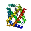
| ||||||||
|---|---|---|---|---|---|---|---|---|---|
| 1 |
| ||||||||
| Unit cell |
|
-Components
| #1: Protein | Mass: 15049.072 Da / Num. of mol.: 1 / Source method: isolated from a natural source / Source: (natural) Glycera dibranchiata (invertebrata) / References: UniProt: P15447 |
|---|---|
| #2: Chemical | ChemComp-HEM /   Mass: 616.487 Da / Num. of mol.: 1 / Source method: obtained synthetically / Formula: C34H32FeN4O4 Mass: 616.487 Da / Num. of mol.: 1 / Source method: obtained synthetically / Formula: C34H32FeN4O4 |
| #3: Water | ChemComp-HOH /  Mass: 18.015 Da / Num. of mol.: 75 / Source method: isolated from a natural source / Formula: H2O Mass: 18.015 Da / Num. of mol.: 75 / Source method: isolated from a natural source / Formula: H2O |
-Experimental details
-Experiment
| Experiment | Method: X-RAY DIFFRACTION / Number of used crystals: 1 |
|---|
- Sample preparation
| Crystal | Density Matthews: 2.57 Å3/Da / Density % sol: 52.05 % | ||||||||||||||||||||||||||||||||||||||||||||||||
|---|---|---|---|---|---|---|---|---|---|---|---|---|---|---|---|---|---|---|---|---|---|---|---|---|---|---|---|---|---|---|---|---|---|---|---|---|---|---|---|---|---|---|---|---|---|---|---|---|---|
| Crystal grow | Temperature: 277 K / Method: vapor diffusion, hanging drop / pH: 4.6 Details: PEG 4000, sodium acetate, ammonium sulfate, pH 4.6, VAPOR DIFFUSION, HANGING DROP, temperature 277K | ||||||||||||||||||||||||||||||||||||||||||||||||
| Crystal grow | *PLUS Method: vapor diffusion | ||||||||||||||||||||||||||||||||||||||||||||||||
| Components of the solutions | *PLUS
|
-Data collection
| Diffraction | Mean temperature: 277 K |
|---|---|
| Diffraction source | Source: ROTATING ANODE / Type: RIGAKU / Wavelength: 1.5418 Å |
| Detector | Type: RIGAKU RAXIS IIC / Detector: IMAGE PLATE / Date: Mar 7, 2000 / Details: mirror |
| Radiation | Monochromator: YALE MIRRORS / Protocol: SINGLE WAVELENGTH / Monochromatic (M) / Laue (L): M / Scattering type: x-ray |
| Radiation wavelength | Wavelength: 1.5418 Å / Relative weight: 1 |
| Reflection | Resolution: 1.4→30 Å / Num. all: 12323 / Num. obs: 11627 / % possible obs: 94.4 % / Observed criterion σ(F): 2 / Observed criterion σ(I): 2 / Redundancy: 3.5 % / Rmerge(I) obs: 0.049 |
| Reflection shell | Resolution: 1.4→30 Å / Redundancy: 3.5 % / % possible all: 90 |
| Reflection | *PLUS Highest resolution: 1.8 Å / Lowest resolution: 10 Å / Num. obs: 10227 / % possible obs: 89 % / Rmerge(I) obs: 0.049 |
| Reflection shell | *PLUS Rmerge(I) obs: 36 |
- Processing
| Software |
| |||||||||||||||||||||||||
|---|---|---|---|---|---|---|---|---|---|---|---|---|---|---|---|---|---|---|---|---|---|---|---|---|---|---|
| Refinement | Method to determine structure: MOLECULAR REPLACEMENT / Resolution: 1.4→10 Å / σ(F): 2 / σ(I): 2
| |||||||||||||||||||||||||
| Refinement step | Cycle: LAST / Resolution: 1.4→10 Å
| |||||||||||||||||||||||||
| Refine LS restraints |
| |||||||||||||||||||||||||
| Refinement | *PLUS Highest resolution: 1.8 Å / Lowest resolution: 10 Å / Num. reflection obs: 10821 / % reflection Rfree: 5 % / Rfactor obs: 0.189 / Rfactor Rfree: 0.241 / Rfactor Rwork: 0.189 | |||||||||||||||||||||||||
| Solvent computation | *PLUS | |||||||||||||||||||||||||
| Displacement parameters | *PLUS | |||||||||||||||||||||||||
| Refine LS restraints | *PLUS
| |||||||||||||||||||||||||
| LS refinement shell | *PLUS Rfactor Rfree: 0.275 / Rfactor Rwork: 0.234 / Rfactor obs: 0.234 |
