 ムービー
ムービー コントローラー


+ データを開く

- 基本情報
基本情報
| 登録情報 | データベース: PDB / ID: 1h21 | |||||||||
|---|---|---|---|---|---|---|---|---|---|---|
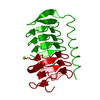
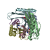
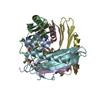
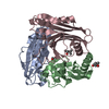
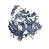
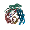
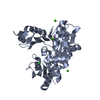
| タイトル | A novel iron centre in the split-Soret cytochrome c from Desulfovibrio desulfuricans ATCC 27774 | |||||||||
 要素 要素 | SPLIT-SORET CYTOCHROME C | |||||||||
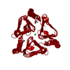
 キーワード キーワード | CYTOCHROME / DIMERIC DI-HEME CYTOCHROME / STACKED HEME ARRANGEMENT / NOVEL FOLD / NOVEL IRON-SULFUR CENTRE / ELECTRON TRANSPORT / SULFATE RESPIRATION | |||||||||
| 機能・相同性 |  機能・相同性情報 機能・相同性情報 | |||||||||
| 生物種 |  DESULFOVIBRIO DESULFURICANS (バクテリア) DESULFOVIBRIO DESULFURICANS (バクテリア) | |||||||||
| 手法 |  X線回折 / シンクロトロン / 多波長異常分散 / 解像度: 2.5 Å X線回折 / シンクロトロン / 多波長異常分散 / 解像度: 2.5 Å | |||||||||
 データ登録者 データ登録者 | Abreu, I.A. / Lourenco, A.I. / Xavier, A.V. / Legall, J. / Coelho, A.V. / Matias, P.M. / Pinto, D.M. / Carrondo, M.A. / Teixeira, M. / Saraiva, L.M. | |||||||||
 引用 引用 | ジャーナル: J.Biol.Inorg.Chem. / 年: 2003 タイトル: A Novel Iron Centre in the Split-Soret Cytochrome C from Desulfovibrio Desulfuricans Atcc 27774 著者: Abreu, I.A. / Lourenco, A.I. / Xavier, A.V. / Legall, J. / Coelho, A.V. / Matias, P.M. / Pinto, D.M. / Armenia Carrondo, M. / Teixeira, M. / Saraiva, L.M. #1: ジャーナル: J.Biol.Inorg.Chem. / 年: 1997タイトル: A Preliminary Analysis of the Three Dimensional Structure of Dimeric Di-Haem Split-Soret Cytochrome C from Desulfovibrio Desulfuricans Atcc 27774 at 2.5 A Resolution Using the MAD ...タイトル: A Preliminary Analysis of the Three Dimensional Structure of Dimeric Di-Haem Split-Soret Cytochrome C from Desulfovibrio Desulfuricans Atcc 27774 at 2.5 A Resolution Using the MAD Phasing Method: A Novel Cytochrome Fold with a Stacked Haem Arrangement 著者: Matias, P.M. / Morais, J. / Coelho, A.V. / Meijers, R. / Gonzalez, A. / Thompson, A.W. / Sieker, L. / Legall, J. / Carrondo, M.A. | |||||||||
| 履歴 |
|
- 構造の表示
構造の表示
| 構造ビューア | 分子: MolmilJmol/JSmol |
|---|
- ダウンロードとリンク
-ダウンロード
| PDBx/mmCIF形式 | 1h21.cif.gz | 202.9 KB | 表示 | PDBx/mmCIF形式 |
|---|---|---|---|---|
| PDB形式 | pdb1h21.ent.gz | 165.6 KB | 表示 | PDB形式 |
| PDBx/mmJSON形式 | 1h21.json.gz | ツリー表示 | PDBx/mmJSON形式 | |
| その他 |  その他のダウンロード その他のダウンロード |
-検証レポート
| アーカイブディレクトリ | https://data.pdbj.org/pub/pdb/validation_reports/h2/1h21ftp://data.pdbj.org/pub/pdb/validation_reports/h2/1h21 | HTTPS FTP |
|---|
-関連構造データ


-リンク
 PDBj
PDBj


- 集合体
集合体
| 登録構造単位 | 
| ||||||||||||
|---|---|---|---|---|---|---|---|---|---|---|---|---|---|
| 1 | 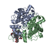
| ||||||||||||
| 2 | 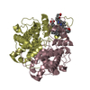
| ||||||||||||
| 単位格子 |
| ||||||||||||
| 非結晶学的対称性 (NCS) | NCS oper:
|
-要素
| #1: タンパク質 | 分子量: 26939.469 Da / 分子数: 4 / 由来タイプ: 天然 由来: (天然) DESULFOVIBRIO DESULFURICANS (バクテリア)参照: UniProt: P81040 #2: 化合物 | ChemComp-HEC /   分子量: 618.503 Da / 分子数: 8 / 由来タイプ: 合成 / 式: C34H34FeN4O4 分子量: 618.503 Da / 分子数: 8 / 由来タイプ: 合成 / 式: C34H34FeN4O4#3: 水 | ChemComp-HOH / |  分子量: 18.015 Da / 分子数: 188 / 由来タイプ: 天然 / 式: H2O 分子量: 18.015 Da / 分子数: 188 / 由来タイプ: 天然 / 式: H2OHas protein modification | Y | 配列の詳細 | GENBANK ENTRY AF465622 IS ON HOLD PENDING MANUSCRIPT ACCEPTANCE FOR PUBLICATION. SEQADV RECORDS ...GENBANK ENTRY AF465622 IS ON HOLD PENDING MANUSCRIPT | |
|---|
-実験情報
-実験
| 実験 | 手法: X線回折 / 使用した結晶の数: 1 |
|---|
- 試料調製
試料調製
| 結晶 | マシュー密度: 2.59 Å3/Da / 溶媒含有率: 53 % | ||||||||||||||||||||||||||||||||||||||||||
|---|---|---|---|---|---|---|---|---|---|---|---|---|---|---|---|---|---|---|---|---|---|---|---|---|---|---|---|---|---|---|---|---|---|---|---|---|---|---|---|---|---|---|---|
| 結晶化 | pH: 5 詳細: PROTEIN WAS CRYSTALLIZED FROM A SOLUTION CONTAINING 0.1-0.2 % (W/V) AGAROSE IN 12-15 % PEG 8K, 0.1M SODIUM ACETATE BUFFER PH 5.0 | ||||||||||||||||||||||||||||||||||||||||||
| 結晶化 | *PLUS 手法: 蒸気拡散法, シッティングドロップ法 / 詳細: Matias, P.M., (1997) J.Biol.Inorg.Chem., 2, 507. | ||||||||||||||||||||||||||||||||||||||||||
| 溶液の組成 | *PLUS
|
-データ収集
| 回折 | 平均測定温度: 100 K |
|---|---|
| 放射光源 | 由来: シンクロトロン / サイト: ESRF  / ビームライン: BM14 / 波長: 1.7401 / ビームライン: BM14 / 波長: 1.7401 |
| 検出器 | タイプ: MARRESEARCH / 検出器: IMAGE PLATE / 日付: 1996年2月15日 / 詳細: MIRRORS |
| 放射 | モノクロメーター: SI (111) / プロトコル: MAD / 単色(M)・ラウエ(L): M / 散乱光タイプ: x-ray |
| 放射波長 | 波長: 1.7401 Å / 相対比: 1 |
| 反射 | 解像度: 2.5→20 Å / Num. obs: 34228 / % possible obs: 93 % / 冗長度: 6.2 % / Rmerge(I) obs: 0.038 / Net I/σ(I): 9.7 |
| 反射 シェル | 解像度: 2.5→2.56 Å / Rmerge(I) obs: 0.074 / Mean I/σ(I) obs: 9.7 / % possible all: 86 |
- 解析
解析
| ソフトウェア |
| ||||||||||||||||||||||||||||||||||||||||||||||||||||||||||||
|---|---|---|---|---|---|---|---|---|---|---|---|---|---|---|---|---|---|---|---|---|---|---|---|---|---|---|---|---|---|---|---|---|---|---|---|---|---|---|---|---|---|---|---|---|---|---|---|---|---|---|---|---|---|---|---|---|---|---|---|---|---|
| 精密化 | 構造決定の手法: 多波長異常分散 / 解像度: 2.5→20 Å / Data cutoff high absF: 100000 / Data cutoff low absF: 0.1 / Isotropic thermal model: RESTRAINED / 交差検証法: THROUGHOUT / σ(F): 2 / 詳細: FIRST 7 RESIDUES NOT VISIBLE IN ELECTRON DENSITY.
| ||||||||||||||||||||||||||||||||||||||||||||||||||||||||||||
| 原子変位パラメータ | Biso mean: 35.04 Å2 | ||||||||||||||||||||||||||||||||||||||||||||||||||||||||||||
| Refine analyze | Luzzati d res low obs: 10 Å / Luzzati sigma a obs: 0.35 Å | ||||||||||||||||||||||||||||||||||||||||||||||||||||||||||||
| 精密化ステップ | サイクル: LAST / 解像度: 2.5→20 Å
| ||||||||||||||||||||||||||||||||||||||||||||||||||||||||||||
| 拘束条件 |
| ||||||||||||||||||||||||||||||||||||||||||||||||||||||||||||
| Xplor file |
| ||||||||||||||||||||||||||||||||||||||||||||||||||||||||||||
| 拘束条件 | *PLUS
|
