[English] 日本語
Yorodumi
- PDB-1gwf: Compound II structure of Micrococcus Lysodeikticus catalase -

+
Open data


ID or keywords:

Loading...

-
Basic information

Entry
Database: PDB / ID: 1gwf
TitleCompound II structure of Micrococcus Lysodeikticus catalase
ComponentsCatalase
KeywordsOXIDOREDUCTASE / CATALASE / HYDROGEN PEROXIDE OXIDOREDUCTASE
Function / homology
Function and homology information


catalase / catalase activity / hydrogen peroxide catabolic process / response to oxidative stress / heme binding / metal ion binding / cytoplasm
Similarity search - Function
Catalase, clade 3 / Catalase, mono-functional, haem-containing, clades 1 and 3 / Catalase HpII, Chain A, domain 1 / Catalase core domain / Catalase haem-binding site / Catalase proximal heme-ligand signature. / Catalase / Catalase active site / Catalase proximal active site signature. / Catalase immune-responsive domain ...Catalase, clade 3 / Catalase, mono-functional, haem-containing, clades 1 and 3 / Catalase HpII, Chain A, domain 1 / Catalase core domain / Catalase haem-binding site / Catalase proximal heme-ligand signature. / Catalase / Catalase active site / Catalase proximal active site signature. / Catalase immune-responsive domain / Catalase-related immune-responsive / Catalase core domain / Catalase, mono-functional, haem-containing / Catalase / catalase family profile. / Catalase superfamily / Beta Barrel / Mainly Beta
Similarity search - Domain/homology
ACETATE ION / PROTOPORPHYRIN IX CONTAINING FE / OXYGEN ATOM / Catalase
Similarity search - Component
Biological speciesMicrococcus luteus (bacteria)
MethodX-RAY DIFFRACTION / OTHER / Resolution: 1.96 Å
AuthorsMurshudov, G.N. / Grebenko, A.I. / Brannigan, J.A. / Antson, A.A. / Barynin, V.V. / Dodson, G.G. / Dauter, Z. / Wilson, K.S. / Melik-Adamyan, W.R.
CitationJournal: Acta Crystallogr.,Sect.D / Year: 2002
Title: The Structures of Micrococcus Lysodeikticus Catalase, its Ferryl Intermediate (Compound II) and Nadph Complex.
Authors: Murshudov, G.N. / Grebenko, A.I. / Brannigan, J.A. / Antson, A.A. / Barynin, V.V. / Dodson, G.G. / Dauter, Z. / Wilson, K.S. / Melik-Adamyan, W.R.
History
DepositionMar 15, 2002Deposition site: PDBE / Processing site: PDBE
Revision 1.0Apr 26, 2002Provider: repository / Type: Initial release
Revision 1.1Apr 1, 2015Group: Database references / Derived calculations ...Database references / Derived calculations / Non-polymer description / Other / Version format compliance
Revision 1.2Jun 20, 2018Group: Data collection / Database references ...Data collection / Database references / Derived calculations / Source and taxonomy / Structure summary
Category: citation / entity ...citation / entity / entity_src_gen / entity_src_nat / pdbx_struct_mod_residue / pdbx_struct_special_symmetry / struct_ref / struct_ref_seq / struct_ref_seq_dif
Item: _citation.page_last / _entity.pdbx_description ..._citation.page_last / _entity.pdbx_description / _entity.src_method / _pdbx_struct_mod_residue.details / _struct_ref.db_code / _struct_ref.pdbx_align_begin / _struct_ref.pdbx_db_accession / _struct_ref.pdbx_seq_one_letter_code / _struct_ref_seq.pdbx_db_accession / _struct_ref_seq_dif.pdbx_seq_db_accession_code
Remark 700 
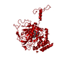
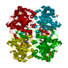
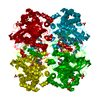
SHEET DETERMINATION METHOD: DSSP THE SHEETS PRESENTED AS "AA" IN EACH CHAIN ON SHEET RECORDS BELOW ... SHEET DETERMINATION METHOD: DSSP THE SHEETS PRESENTED AS "AA" IN EACH CHAIN ON SHEET RECORDS BELOW IS ACTUALLY AN 11-STRANDED BARREL THIS IS REPRESENTED BY A 12-STRANDED SHEET IN WHICH THE FIRST AND LAST STRANDS ARE IDENTICAL.

-
Structure visualization

Structure viewerMolecule:
MolmilJmol/JSmol

Downloads & links

-
Assembly

Deposited unit
A: Catalase
hetero molecules


Theoretical massNumber of molelcules
Total (without water)58,0818
Polymers57,0431
Non-polymers1,0397
Water11,223623
1
A: Catalase
hetero molecules

A: Catalase
hetero molecules

A: Catalase
hetero molecules

A: Catalase
hetero molecules


Theoretical massNumber of molelcules
Total (without water)232,32532
Polymers228,1704
Non-polymers4,15528
Water724
TypeNameSymmetry operationNumber
identity operation1_555x,y,z1
crystal symmetry operation2_555-x,-y,z1
crystal symmetry operation8_555-y,-x,-z1
crystal symmetry operation7_555y,x,-z1
MethodPQS
Unit cell
Length a, b, c (Å)106.190, 106.190, 105.300
Angle α, β, γ (deg.)90.00, 90.00, 90.00
Int Tables number94
Space group name H-MP42212
Components on special symmetry positions
IDModelComponents
11A-2062-

HOH

-
Components

-
Protein , 1 types, 1 molecules A

#1: Protein Catalase /


Mass: 57042.582 Da / Num. of mol.: 1
Source method: isolated from a genetically manipulated source
Source: (gene. exp.) Micrococcus luteus (bacteria) / Gene: katA / Production host: Escherichia coli (E. coli) / References: UniProt: P29422, catalase

-
Non-polymers , 5 types, 630 molecules

#2: Chemical ChemComp-HEM / PROTOPORPHYRIN IX CONTAINING FE / HEME / Heme B


Mass: 616.487 Da / Num. of mol.: 1 / Source method: obtained synthetically / Formula: C34H32FeN4O4
#3: Chemical ChemComp-SO4 / SULFATE ION / Sulfate


Mass: 96.063 Da / Num. of mol.: 3 / Source method: obtained synthetically / Formula: SO4
#4: Chemical ChemComp-ACT / ACETATE ION / Acetate


Mass: 59.044 Da / Num. of mol.: 2 / Source method: obtained synthetically / Formula: C2H3O2
#5: Chemical ChemComp-O / OXYGEN ATOM / Oxygen


Mass: 15.999 Da / Num. of mol.: 1 / Source method: obtained synthetically / Formula: O
#6: Water ChemComp-HOH / water / Water


Mass: 18.015 Da / Num. of mol.: 623 / Source method: isolated from a natural source / Formula: H2O

-
Experimental details

-
Experiment

ExperimentMethod: X-RAY DIFFRACTION / Number of used crystals: 1

-
Sample preparation

CrystalDensity Matthews: 2.6 Å3/Da / Density % sol: 52.73 %
Crystal growpH: 5.2 / Details: pH 5.20

-
Data collection

DiffractionMean temperature: 120 K
Diffraction sourceSource: ROTATING ANODE / Type: RIGAKU / Wavelength: 1.54
DetectorType: MARRESEARCH / Detector: IMAGE PLATE / Date: Oct 15, 1995
RadiationProtocol: SINGLE WAVELENGTH / Monochromatic (M) / Laue (L): M / Scattering type: x-ray
Radiation wavelengthWavelength: 1.54 Å / Relative weight: 1
ReflectionResolution: 1.936→74.536 Å / Num. obs: 41376 / % possible obs: 99.5 % / Redundancy: 5 % / Rmerge(I) obs: 0.061 / Net I/σ(I): 8

-
Processing

Software
NameVersionClassification
REFMAC5refinement
DENZOdata reduction
SCALEPACKdata scaling
RefinementMethod to determine structure: OTHER / Resolution: 1.96→74.54 Å / SU B: 2.755 / SU ML: 0.079 / Cross valid method: THROUGHOUT / ESU R Free: 0.128 / Details: NONE
RfactorNum. reflection% reflectionSelection details
Rfree0.18966 2189 5 %RANDOM
Rwork0.14095 ---
obs0.08935 41376 99.15 %-
Refinement stepCycle: LAST / Resolution: 1.96→74.54 Å
ProteinNucleic acidLigandSolventTotal
Num. atoms3986 0 67 623 4676

+
About Yorodumi

-
News

-
Feb 9, 2022. New format data for meta-information of EMDB entries

New format data for meta-information of EMDB entries

  • Version 3 of the EMDB header file is now the official format.
  • The previous official version 1.9 will be removed from the archive.

Related info.:EMDB header

External links:wwPDB to switch to version 3 of the EMDB data model

-
Aug 12, 2020. Covid-19 info

Covid-19 info

URL: https://pdbj.org/emnavi/covid19.php

New page: Covid-19 featured information page in EM Navigator.

Related info.:Covid-19 info / Mar 5, 2020. Novel coronavirus structure data

+
Mar 5, 2020. Novel coronavirus structure data

Novel coronavirus structure data

Related info.:Yorodumi Speices / Aug 12, 2020. Covid-19 info

External links:COVID-19 featured content - PDBj / Molecule of the Month (242):Coronavirus Proteases

+
Jan 31, 2019. EMDB accession codes are about to change! (news from PDBe EMDB page)

EMDB accession codes are about to change! (news from PDBe EMDB page)

  • The allocation of 4 digits for EMDB accession codes will soon come to an end. Whilst these codes will remain in use, new EMDB accession codes will include an additional digit and will expand incrementally as the available range of codes is exhausted. The current 4-digit format prefixed with “EMD-” (i.e. EMD-XXXX) will advance to a 5-digit format (i.e. EMD-XXXXX), and so on. It is currently estimated that the 4-digit codes will be depleted around Spring 2019, at which point the 5-digit format will come into force.
  • The EM Navigator/Yorodumi systems omit the EMD- prefix.

Related info.:Q: What is EMD? / ID/Accession-code notation in Yorodumi/EM Navigator

External links:EMDB Accession Codes are Changing Soon! / Contact to PDBj

+
Jul 12, 2017. Major update of PDB

Major update of PDB

  • wwPDB released updated PDB data conforming to the new PDBx/mmCIF dictionary.
  • This is a major update changing the version number from 4 to 5, and with Remediation, in which all the entries are updated.
  • In this update, many items about electron microscopy experimental information are reorganized (e.g. em_software).
  • Now, EM Navigator and Yorodumi are based on the updated data.

External links:wwPDB Remediation / Enriched Model Files Conforming to OneDep Data Standards Now Available in the PDB FTP Archive

-
Yorodumi

Thousand views of thousand structures

  • Yorodumi is a browser for structure data from EMDB, PDB, SASBDB, etc.
  • This page is also the successor to EM Navigator detail page, and also detail information page/front-end page for Omokage search.
  • The word "yorodu" (or yorozu) is an old Japanese word meaning "ten thousand". "mi" (miru) is to see.

Related info.:EMDB / PDB / SASBDB / Comparison of 3 databanks / Yorodumi Search / Aug 31, 2016. New EM Navigator & Yorodumi / Yorodumi Papers / Jmol/JSmol / Function and homology information / Changes in new EM Navigator and Yorodumi

Read more