 Movie
Movie Controller

[English] 日本語
 Yorodumi
- PDB-1gv3: The 2.0 Angstrom resolution structure of the catalytic portion of... -
+ Open data

- Basic information
| Entry | Database: PDB / ID: 1gv3 | ||||||
|---|---|---|---|---|---|---|---|
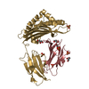
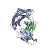
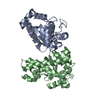
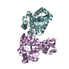
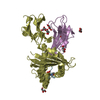
| Title | The 2.0 Angstrom resolution structure of the catalytic portion of a cyanobacterial membrane-bound manganese superoxide dismutase | ||||||
 Components Components | MANGANESE SUPEROXIDE DISMUTASE | ||||||
 Keywords Keywords | MANGANESE SUPEROXIDE DISMUTASE / ANABAENA PCC 7120 | ||||||
| Function / homology |  Function and homology information Function and homology informationsuperoxide dismutase / superoxide dismutase activity / metal ion binding / cytoplasm Similarity search - Function | ||||||
| Biological species |  ANABAENA SP. (bacteria) ANABAENA SP. (bacteria) | ||||||
| Method |  X-RAY DIFFRACTION / MOLECULAR REPLACEMENT / Resolution: 2 Å X-RAY DIFFRACTION / MOLECULAR REPLACEMENT / Resolution: 2 Å | ||||||
 Authors Authors | Atzenhofer, W. / Regelsberger, G. / Jacob, U. / Huber, R. / Peschek, G.A. / Obinger, C. | ||||||
 Citation Citation | Journal: J.Mol.Biol. / Year: 2002 Title: The 2.0A Resolution Structure of the Catalytic Portion of a Cyanobacterial Membrane-Bound Manganese Superoxide Dismutase Authors: Atzenhofer, W. / Regelsberger, G. / Jacob, U. / Peschek, G.A. / Furtmuller, P. / Huber, R. / Obinger, C. | ||||||
| History |
|
- Structure visualization
| Structure viewer | Molecule: MolmilJmol/JSmol |
|---|
- Downloads & links
-Download
| PDBx/mmCIF format | 1gv3.cif.gz | 100.7 KB | Display | PDBx/mmCIF format |
|---|---|---|---|---|
| PDB format | pdb1gv3.ent.gz | 77 KB | Display | PDB format |
| PDBx/mmJSON format | 1gv3.json.gz | Tree view | PDBx/mmJSON format | |
| Others |  Other downloads Other downloads |
-Validation report
| Arichive directory | https://data.pdbj.org/pub/pdb/validation_reports/gv/1gv3ftp://data.pdbj.org/pub/pdb/validation_reports/gv/1gv3 | HTTPS FTP |
|---|
-Related structure data
| Related structure data |  1mngS S: Starting model for refinement |
|---|---|
| Similar structure data |


-Links
 PDBj
PDBj


- Assembly
| Deposited unit | 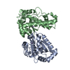
| ||||||||
|---|---|---|---|---|---|---|---|---|---|
| 1 |
| ||||||||
| Unit cell |
|
-Components
| #1: Protein | Mass: 28213.043 Da / Num. of mol.: 2 Fragment: HELICAL HAIRPIN, ALPHA/BETA DOMAIN, RESIDUES 30-270 Source method: isolated from a genetically manipulated source Source: (gene. exp.) ANABAENA SP. (bacteria) / Strain: PCC 7120 / Plasmid: PET-3A / Production host: #2: Chemical |   Mass: 54.938 Da / Num. of mol.: 2 / Source method: obtained synthetically / Formula: Mn Mass: 54.938 Da / Num. of mol.: 2 / Source method: obtained synthetically / Formula: Mn#3: Water | ChemComp-HOH / |  Mass: 18.015 Da / Num. of mol.: 166 / Source method: isolated from a natural source / Formula: H2O Mass: 18.015 Da / Num. of mol.: 166 / Source method: isolated from a natural source / Formula: H2O |
|---|
-Experimental details
-Experiment
| Experiment | Method: X-RAY DIFFRACTION / Number of used crystals: 3 |
|---|
- Sample preparation
| Crystal | Density Matthews: 2.71 Å3/Da / Density % sol: 54.53 % | ||||||||||||||||||||||||||||||
|---|---|---|---|---|---|---|---|---|---|---|---|---|---|---|---|---|---|---|---|---|---|---|---|---|---|---|---|---|---|---|---|
| Crystal grow | pH: 7.5 / Details: pH 7.50 | ||||||||||||||||||||||||||||||
| Crystal grow | *PLUS Method: vapor diffusion, sitting drop | ||||||||||||||||||||||||||||||
| Components of the solutions | *PLUS
|
-Data collection
| Diffraction | Mean temperature: 287 K |
|---|---|
| Diffraction source | Source: ROTATING ANODE / Type: RIGAKU / Wavelength: 1.518 |
| Detector | Type: MARRESEARCH / Detector: IMAGE PLATE / Date: Aug 15, 2001 |
| Radiation | Monochromator: GRAPHIT / Protocol: SINGLE WAVELENGTH / Monochromatic (M) / Laue (L): M / Scattering type: x-ray |
| Radiation wavelength | Wavelength: 1.518 Å / Relative weight: 1 |
| Reflection | Resolution: 2.03→30.57 Å / Num. obs: 35333 / % possible obs: 93.9 % / Redundancy: 2.5 % |
| Reflection | *PLUS Num. measured all: 124807 |
- Processing
| Software |
| ||||||||||||||||||||||||||||||||||||||||||||||||||||||||||||||||||||||||||||||||
|---|---|---|---|---|---|---|---|---|---|---|---|---|---|---|---|---|---|---|---|---|---|---|---|---|---|---|---|---|---|---|---|---|---|---|---|---|---|---|---|---|---|---|---|---|---|---|---|---|---|---|---|---|---|---|---|---|---|---|---|---|---|---|---|---|---|---|---|---|---|---|---|---|---|---|---|---|---|---|---|---|---|
| Refinement | Method to determine structure: MOLECULAR REPLACEMENT Starting model: 1MNG Resolution: 2→100 Å / Data cutoff high absF: 10000 / σ(F): 0
| ||||||||||||||||||||||||||||||||||||||||||||||||||||||||||||||||||||||||||||||||
| Displacement parameters |
| ||||||||||||||||||||||||||||||||||||||||||||||||||||||||||||||||||||||||||||||||
| Refinement step | Cycle: LAST / Resolution: 2→100 Å
| ||||||||||||||||||||||||||||||||||||||||||||||||||||||||||||||||||||||||||||||||
| Refine LS restraints |
| ||||||||||||||||||||||||||||||||||||||||||||||||||||||||||||||||||||||||||||||||
| Refinement | *PLUS Highest resolution: 2.03 Å / Lowest resolution: 30.57 Å | ||||||||||||||||||||||||||||||||||||||||||||||||||||||||||||||||||||||||||||||||
| Solvent computation | *PLUS | ||||||||||||||||||||||||||||||||||||||||||||||||||||||||||||||||||||||||||||||||
| Displacement parameters | *PLUS |
