 Movie
Movie Controller


+ Open data

- Basic information
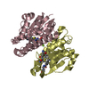
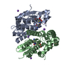
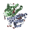
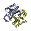
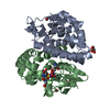
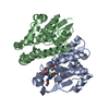
| Entry | Database: PDB / ID: 1gnw | ||||||
|---|---|---|---|---|---|---|---|
| Title | STRUCTURE OF GLUTATHIONE S-TRANSFERASE | ||||||
 Components Components | GLUTATHIONE S-TRANSFERASE | ||||||
 Keywords Keywords | TRANSFERASE / HERBICIDE DETOXIFICATION | ||||||
| Function / homology |  Function and homology information Function and homology informationresponse to oomycetes / camalexin binding / quercitrin binding / salicylic acid binding / toxin catabolic process / auxin-activated signaling pathway / glutathione binding / plasmodesma / apoplast / plant-type vacuole ...response to oomycetes / camalexin binding / quercitrin binding / salicylic acid binding / toxin catabolic process / auxin-activated signaling pathway / glutathione binding / plasmodesma / apoplast / plant-type vacuole / chloroplast stroma / response to zinc ion / glutathione transferase / glutathione transferase activity / response to cadmium ion / response to cold / chloroplast / peroxidase activity / defense response / protein domain specific binding / intracellular membrane-bounded organelle / endoplasmic reticulum / plasma membrane / cytosol Similarity search - Function | ||||||
| Biological species |  | ||||||
| Method |  X-RAY DIFFRACTION / Resolution: 2.2 Å X-RAY DIFFRACTION / Resolution: 2.2 Å | ||||||
 Authors Authors | Reinemer, P. / Prade, L. / Hof, P. / Neuefeind, T. / Huber, R. / Palme, K. / Bartunik, H.D. / Bieseler, B. | ||||||
 Citation Citation | Journal: J.Mol.Biol. / Year: 1996 Title: Three-dimensional structure of glutathione S-transferase from Arabidopsis thaliana at 2.2 A resolution: structural characterization of herbicide-conjugating plant glutathione S-transferases ...Title: Three-dimensional structure of glutathione S-transferase from Arabidopsis thaliana at 2.2 A resolution: structural characterization of herbicide-conjugating plant glutathione S-transferases and a novel active site architecture. Authors: Reinemer, P. / Prade, L. / Hof, P. / Neuefeind, T. / Huber, R. / Zettl, R. / Palme, K. / Schell, J. / Koelln, I. / Bartunik, H.D. / Bieseler, B. | ||||||
| History |
|
- Structure visualization
| Structure viewer | Molecule: MolmilJmol/JSmol |
|---|
- Downloads & links
-Download
| PDBx/mmCIF format | 1gnw.cif.gz | 124.3 KB | Display | PDBx/mmCIF format |
|---|---|---|---|---|
| PDB format | pdb1gnw.ent.gz | 99.5 KB | Display | PDB format |
| PDBx/mmJSON format | 1gnw.json.gz | Tree view | PDBx/mmJSON format | |
| Others |  Other downloads Other downloads |
-Validation report
| Summary document | 1gnw_validation.pdf.gz | 1.1 MB | Display | wwPDB validaton report |
|---|---|---|---|---|
| Full document | 1gnw_full_validation.pdf.gz | 1.1 MB | Display | |
| Data in XML | 1gnw_validation.xml.gz | 21.3 KB | Display | |
| Data in CIF | 1gnw_validation.cif.gz | 29.7 KB | Display | |
| Arichive directory | https://data.pdbj.org/pub/pdb/validation_reports/gn/1gnwftp://data.pdbj.org/pub/pdb/validation_reports/gn/1gnw | HTTPS FTP |
-Related structure data
| Similar structure data |
|---|


-Links
 PDBj
PDBj

- Assembly
| Deposited unit | 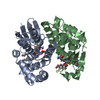
| ||||||||
|---|---|---|---|---|---|---|---|---|---|
| 1 |
| ||||||||
| Unit cell |
|
-Components
| #1: Protein | Mass: 24031.371 Da / Num. of mol.: 2 Source method: isolated from a genetically manipulated source Source: (gene. exp.)  #2: Chemical | ChemComp-GTX / 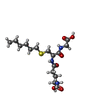  Mass: 392.491 Da / Num. of mol.: 4 / Source method: obtained synthetically / Formula: C16H30N3O6S Mass: 392.491 Da / Num. of mol.: 4 / Source method: obtained synthetically / Formula: C16H30N3O6S#3: Water | ChemComp-HOH / |  Mass: 18.015 Da / Num. of mol.: 220 / Source method: isolated from a natural source / Formula: H2O Mass: 18.015 Da / Num. of mol.: 220 / Source method: isolated from a natural source / Formula: H2O |
|---|
-Experimental details
-Experiment
| Experiment | Method: X-RAY DIFFRACTION |
|---|
- Sample preparation
| Crystal | Density Matthews: 2.7 Å3/Da / Density % sol: 54.38 % | ||||||||||||||||||||||||||||||||||||||||||||||||||||||||||||||||||
|---|---|---|---|---|---|---|---|---|---|---|---|---|---|---|---|---|---|---|---|---|---|---|---|---|---|---|---|---|---|---|---|---|---|---|---|---|---|---|---|---|---|---|---|---|---|---|---|---|---|---|---|---|---|---|---|---|---|---|---|---|---|---|---|---|---|---|---|
| Crystal grow | *PLUS pH: 7 / Method: vapor diffusion, sitting drop | ||||||||||||||||||||||||||||||||||||||||||||||||||||||||||||||||||
| Components of the solutions | *PLUS
|
-Data collection
| Diffraction source | Wavelength: 1.5418 |
|---|---|
| Detector | Type: ENRAF-NONIUS FAST / Detector: DIFFRACTOMETER / Date: 1994 |
| Radiation | Monochromatic (M) / Laue (L): M / Scattering type: x-ray |
| Radiation wavelength | Wavelength: 1.5418 Å / Relative weight: 1 |
| Reflection | Num. obs: 23438 / % possible obs: 88.9 % / Redundancy: 4.6 % / Rmerge(I) obs: 0.094 |
| Reflection | *PLUS Highest resolution: 2.2 Å / Lowest resolution: 9999 Å |
| Reflection shell | *PLUS Highest resolution: 2.2 Å / Lowest resolution: 2.23 Å / % possible obs: 46.2 % |
- Processing
| Software |
| ||||||||||||||||||||||||||||||||||||||||||||||||||||||||||||
|---|---|---|---|---|---|---|---|---|---|---|---|---|---|---|---|---|---|---|---|---|---|---|---|---|---|---|---|---|---|---|---|---|---|---|---|---|---|---|---|---|---|---|---|---|---|---|---|---|---|---|---|---|---|---|---|---|---|---|---|---|---|
| Refinement | Resolution: 2.2→8 Å /
| ||||||||||||||||||||||||||||||||||||||||||||||||||||||||||||
| Displacement parameters | Biso mean: 19.5 Å2 | ||||||||||||||||||||||||||||||||||||||||||||||||||||||||||||
| Refine analyze | Luzzati coordinate error obs: 0.19 Å | ||||||||||||||||||||||||||||||||||||||||||||||||||||||||||||
| Refinement step | Cycle: LAST / Resolution: 2.2→8 Å
| ||||||||||||||||||||||||||||||||||||||||||||||||||||||||||||
| Refine LS restraints |
| ||||||||||||||||||||||||||||||||||||||||||||||||||||||||||||
| Software | *PLUS Name: X-PLOR / Classification: refinement | ||||||||||||||||||||||||||||||||||||||||||||||||||||||||||||
| Refinement | *PLUS | ||||||||||||||||||||||||||||||||||||||||||||||||||||||||||||
| Solvent computation | *PLUS | ||||||||||||||||||||||||||||||||||||||||||||||||||||||||||||
| Displacement parameters | *PLUS | ||||||||||||||||||||||||||||||||||||||||||||||||||||||||||||
| Refine LS restraints | *PLUS
| ||||||||||||||||||||||||||||||||||||||||||||||||||||||||||||
| LS refinement shell | *PLUS Highest resolution: 2.2 Å / Lowest resolution: 2.22 Å / Rfactor obs: 0.289 |
