 Movie
Movie Controller

[English] 日本語
 Yorodumi
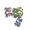
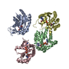
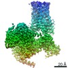
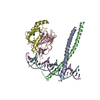
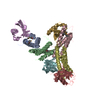
- PDB-1gdt: CRYSTAL STRUCTURE OF A SITE-SPECIFIC RECOMBINASE, GAMMA-DELTA RES... -
+ Open data

- Basic information
| Entry | Database: PDB / ID: 1gdt | ||||||
|---|---|---|---|---|---|---|---|
| Title | CRYSTAL STRUCTURE OF A SITE-SPECIFIC RECOMBINASE, GAMMA-DELTA RESOLVASE COMPLEXED WITH A 34 BP CLEAVAGE SITE | ||||||
 Components Components |
| ||||||
 Keywords Keywords | DNA BINDING PROTEIN/DNA /  PROTEIN-DNA COMPLEX / DOUBLE HELIX / OVERHANGING BASE / DNA BINDING PROTEIN-DNA COMPLEX PROTEIN-DNA COMPLEX / DOUBLE HELIX / OVERHANGING BASE / DNA BINDING PROTEIN-DNA COMPLEX | ||||||
| Function / homology |  Function and homology information Function and homology information | ||||||
| Biological species |  Escherichia coli (E. coli) Escherichia coli (E. coli) | ||||||
| Method | X-RAY DIFFRACTION / Resolution: 3 Å | ||||||
 Authors Authors | Yang, W. / Steitz, T.A. | ||||||
 Citation Citation | Journal: Cell(Cambridge,Mass.) / Year: 1995 Title: Crystal structure of the site-specific recombinase gamma delta resolvase complexed with a 34 bp cleavage site. Authors: Yang, W. / Steitz, T.A. | ||||||
| History |
|
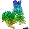
- Structure visualization
| Structure viewer | Molecule: MolmilJmol/JSmol |
|---|
- Downloads & links
-Download
| PDBx/mmCIF format | 1gdt.cif.gz | 118.2 KB | Display | PDBx/mmCIF format |
|---|---|---|---|---|
| PDB format | pdb1gdt.ent.gz | 89.4 KB | Display | PDB format |
| PDBx/mmJSON format | 1gdt.json.gz | Tree view | PDBx/mmJSON format | |
| Others |  Other downloads Other downloads |
-Validation report
| Arichive directory | https://data.pdbj.org/pub/pdb/validation_reports/gd/1gdtftp://data.pdbj.org/pub/pdb/validation_reports/gd/1gdt | HTTPS FTP |
|---|
-Related structure data
| Similar structure data |
|---|


-Links
 PDBj
PDBj


- Assembly
| Deposited unit | 
| ||||||||
|---|---|---|---|---|---|---|---|---|---|
| 1 |
| ||||||||
| Unit cell |
|
-Components
| #1: DNA chain | Mass: 6774.416 Da / Num. of mol.: 1 / Source method: obtained synthetically | ||||||||
|---|---|---|---|---|---|---|---|---|---|
| #2: DNA chain | Mass: 3966.597 Da / Num. of mol.: 2 / Source method: obtained synthetically #3: DNA chain | | Mass: 6445.209 Da / Num. of mol.: 1 / Source method: obtained synthetically #4: Protein | Mass: 20394.465 Da / Num. of mol.: 2 Source method: isolated from a genetically manipulated source Source: (gene. exp.) Escherichia coli (E. coli) / Strain: STRAINS THAT CONTAIN F EPISOME / Gene: TNPR / Plasmid: MGH285 / Gene (production host): TNPR / Production host: Escherichia coli (E. coli) / Strain (production host): AR120 / References: UniProt: P03012#5: Water | ChemComp-HOH / | Water Mass: 18.015 Da / Num. of mol.: 29 / Source method: isolated from a natural source / Formula: H2O Mass: 18.015 Da / Num. of mol.: 29 / Source method: isolated from a natural source / Formula: H2OCompound details | RESOLVASE GENE IS PUT UNDER THE CONTROL OF THE LAMBDA PROMOTER AND INDUCED BY NALIDIXIC ACID. | |
-Experimental details
-Experiment
| Experiment | Method: X-RAY DIFFRACTION |
|---|
- Sample preparation
| Crystal | Density Matthews: 2.64 Å3/Da / Density % sol: 54 % | |||||||||||||||||||||||||||||||||||||||||||||||||||||||||||||||||
|---|---|---|---|---|---|---|---|---|---|---|---|---|---|---|---|---|---|---|---|---|---|---|---|---|---|---|---|---|---|---|---|---|---|---|---|---|---|---|---|---|---|---|---|---|---|---|---|---|---|---|---|---|---|---|---|---|---|---|---|---|---|---|---|---|---|---|
| Crystal | *PLUS Density % sol: 54 % | |||||||||||||||||||||||||||||||||||||||||||||||||||||||||||||||||
| Crystal grow | *PLUS pH: 6 / Method: vapor diffusion, hanging drop | |||||||||||||||||||||||||||||||||||||||||||||||||||||||||||||||||
| Components of the solutions | *PLUS
|
-Data collection
| Diffraction | Mean temperature: 113 K |
|---|---|
| Detector | Date: Apr 1, 1994 |
| Radiation | Protocol: SINGLE WAVELENGTH / Monochromatic (M) / Laue (L): M / Scattering type: x-ray |
| Radiation wavelength | Relative weight: 1 |
| Reflection | Resolution: 3→30 Å / Num. obs: 13255 / % possible obs: 94.2 % / Observed criterion σ(I): 0 / Rmerge(I) obs: 0.077 |
| Reflection | *PLUS Highest resolution: 3 Å / Lowest resolution: 30 Å / % possible obs: 94.2 % / Observed criterion σ(I): 0 |
- Processing
| Software |
| ||||||||||||||||||||||||||||||||||||||||||||||||||||||||||||
|---|---|---|---|---|---|---|---|---|---|---|---|---|---|---|---|---|---|---|---|---|---|---|---|---|---|---|---|---|---|---|---|---|---|---|---|---|---|---|---|---|---|---|---|---|---|---|---|---|---|---|---|---|---|---|---|---|---|---|---|---|---|
| Refinement | Resolution: 3→10 Å / σ(F): 2
| ||||||||||||||||||||||||||||||||||||||||||||||||||||||||||||
| Displacement parameters | Biso mean: 26.69 Å2 | ||||||||||||||||||||||||||||||||||||||||||||||||||||||||||||
| Refine analyze | Luzzati coordinate error obs: 0.4 Å | ||||||||||||||||||||||||||||||||||||||||||||||||||||||||||||
| Refinement step | Cycle: LAST / Resolution: 3→10 Å
| ||||||||||||||||||||||||||||||||||||||||||||||||||||||||||||
| Refine LS restraints |
| ||||||||||||||||||||||||||||||||||||||||||||||||||||||||||||
| Software | *PLUS Name: X-PLOR / Classification: refinement | ||||||||||||||||||||||||||||||||||||||||||||||||||||||||||||
| Refinement | *PLUS Highest resolution: 3 Å / Lowest resolution: 10 Å / σ(F): 2 | ||||||||||||||||||||||||||||||||||||||||||||||||||||||||||||
| Solvent computation | *PLUS | ||||||||||||||||||||||||||||||||||||||||||||||||||||||||||||
| Displacement parameters | *PLUS | ||||||||||||||||||||||||||||||||||||||||||||||||||||||||||||
| Refine LS restraints | *PLUS
|
