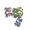
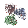
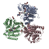
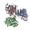
Entry Database : PDB / ID : 4cl1Title The crystal structure of NS5A domain 1 from genotype 1a reveals new clues to the mechanism of action for dimeric HCV inhibitors NON-STRUCTURAL PROTEIN 5A Keywords Function / homology Function Domain/homology Component
/ / / / / / / / / / / / / / / / / / / / / / / / / / / / / / / / / / / / / / / / / / / / / / / / / / / / / / / / / / / / / / / / / / / / / / / / / / / / / / / / / / / / / / / / / / / / / / / / / / / / / / / / / / / / Biological species Method / / / Resolution : 3.5 Å Authors Lambert, S.M. / Langley, D.R. / Garnett, J.A. / Angell, R. / Hedgethorne, K. / Meanwell, N.A. / Matthews, S.J. Journal : Protein Sci. / Year : 2014Title : The Crystal Structure of Ns5A Domain 1 from Genotype 1A Reveals New Clues to the Mechanism of Action for Dimeric Hcv Inhibitors.Authors : Lambert, S.M. / Langley, D.R. / Garnett, J.A. / Angell, R. / Hedgethorne, K. / Meanwell, N.A. / Matthews, S.J. History Deposition Jan 10, 2014 Deposition site / Processing site Revision 1.0 Apr 2, 2014 Provider / Type Revision 1.1 Jun 4, 2014 Group Revision 1.2 Jun 20, 2018 Group / Data collection / Derived calculationsCategory database_PDB_caveat / pdbx_struct_conn_angle ... database_PDB_caveat / pdbx_struct_conn_angle / pdbx_validate_close_contact / pdbx_validate_symm_contact / struct_conn Item _pdbx_struct_conn_angle.ptnr1_auth_seq_id / _pdbx_struct_conn_angle.ptnr1_label_seq_id ... _pdbx_struct_conn_angle.ptnr1_auth_seq_id / _pdbx_struct_conn_angle.ptnr1_label_seq_id / _pdbx_struct_conn_angle.ptnr3_auth_seq_id / _pdbx_struct_conn_angle.ptnr3_label_seq_id / _pdbx_struct_conn_angle.value Revision 1.3 Feb 20, 2019 Group / Data collection / Derived calculationsCategory pdbx_data_processing_status / pdbx_seq_map_depositor_info ... pdbx_data_processing_status / pdbx_seq_map_depositor_info / pdbx_validate_close_contact / pdbx_validate_symm_contact / struct_biol / struct_conn / struct_conn_type Item / _pdbx_seq_map_depositor_info.one_letter_code_modRevision 1.4 Dec 20, 2023 Group Data collection / Database references ... Data collection / Database references / Derived calculations / Other / Refinement description Category chem_comp_atom / chem_comp_bond ... chem_comp_atom / chem_comp_bond / database_2 / pdbx_database_status / pdbx_initial_refinement_model / struct_site Item _database_2.pdbx_DOI / _database_2.pdbx_database_accession ... _database_2.pdbx_DOI / _database_2.pdbx_database_accession / _pdbx_database_status.status_code_sf / _struct_site.pdbx_auth_asym_id / _struct_site.pdbx_auth_comp_id / _struct_site.pdbx_auth_seq_id Revision 1.5 Nov 20, 2024 Group / Category / pdbx_modification_feature
Show all Show less
 Movie
Movie Controller

 Yorodumi
 Open data
 Basic information
 Components
 Keywords
 Function and homology information
 HEPATITIS C VIRUS
 X-RAY DIFFRACTION /
  Authors
 Citation
 Structure visualization
 Downloads & links
 Other downloads


 PDBj
PDBj
 Assembly


 Mass: 65.409 Da / Num. of mol.: 4 / Source method: obtained synthetically / Formula: Zn
 Mass: 96.063 Da / Num. of mol.: 1 / Source method: obtained synthetically / Formula: SO4
 Sample preparation
 / Beamline: I24 / Wavelength: 0.9778
  Processing