 Movie
Movie Controller

[English] 日本語
 Yorodumi
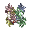
- PDB-1f9n: CRYSTAL STRUCTURE OF AHRC, THE ARGININE REPRESSOR/ACTIVATOR PROTE... -
+ Open data

- Basic information
| Entry | Database: PDB / ID: 1f9n | ||||||
|---|---|---|---|---|---|---|---|
| Title | CRYSTAL STRUCTURE OF AHRC, THE ARGININE REPRESSOR/ACTIVATOR PROTEIN FROM BACILLUS SUBTILIS | ||||||
 Components Components | ARGININE REPRESSOR/ACTIVATOR PROTEIN | ||||||
 Keywords Keywords | DNA BINDING PROTEIN / WINGED-HELIX-TURN-HELIX / ARGININE REPRESSOR | ||||||
| Function / homology |  Function and homology information Function and homology informationregulation of arginine biosynthetic process / : / L-arginine biosynthetic process / arginine binding / protein complex oligomerization / cis-regulatory region sequence-specific DNA binding / transcription regulator complex / DNA-binding transcription factor activity / cytoplasm Similarity search - Function | ||||||
| Biological species |  | ||||||
| Method |  X-RAY DIFFRACTION / SYNCHROTRON / Resolution: 2.7 Å X-RAY DIFFRACTION / SYNCHROTRON / Resolution: 2.7 Å | ||||||
 Authors Authors | Dennis, C.A. / Glykos, N.M. / Parsons, M.R. / Phillips, S.E.V. | ||||||
 Citation Citation | Journal: Acta Crystallogr.,Sect.D / Year: 2002 Title: The structure of AhrC, the arginine repressor/activator protein from Bacillus subtilis. Authors: Dennis C, C.A. / Glykos, N.M. / Parsons, M.R. / Phillips, S.E. | ||||||
| History |
|
- Structure visualization
| Structure viewer | Molecule: MolmilJmol/JSmol |
|---|
- Downloads & links
-Download
| PDBx/mmCIF format | 1f9n.cif.gz | 180.6 KB | Display | PDBx/mmCIF format |
|---|---|---|---|---|
| PDB format | pdb1f9n.ent.gz | 147 KB | Display | PDB format |
| PDBx/mmJSON format | 1f9n.json.gz | Tree view | PDBx/mmJSON format | |
| Others |  Other downloads Other downloads |
-Validation report
| Arichive directory | https://data.pdbj.org/pub/pdb/validation_reports/f9/1f9nftp://data.pdbj.org/pub/pdb/validation_reports/f9/1f9n | HTTPS FTP |
|---|
-Related structure data
| Similar structure data |
|---|


-Links
 PDBj
PDBj- Assembly
| Deposited unit | 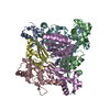
| ||||||||
|---|---|---|---|---|---|---|---|---|---|
| 1 |
| ||||||||
| Unit cell |
| ||||||||
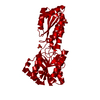
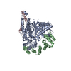
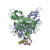
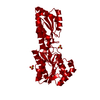
| Details | The biological assembly is a hexamer with 32 symmetry. The hexamer is formed by a dimer of trimers. |
-Components
| #1: Protein | Mass: 16854.479 Da / Num. of mol.: 6 Source method: isolated from a genetically manipulated source Source: (gene. exp.) #2: Water | ChemComp-HOH / |  Mass: 18.015 Da / Num. of mol.: 240 / Source method: isolated from a natural source / Formula: H2O Mass: 18.015 Da / Num. of mol.: 240 / Source method: isolated from a natural source / Formula: H2O |
|---|
-Experimental details
-Experiment
| Experiment | Method: X-RAY DIFFRACTION / Number of used crystals: 1 |
|---|
- Sample preparation
| Crystal | Density Matthews: 2.92 Å3/Da / Density % sol: 57.87 % | ||||||||||||||||||||||||||||||
|---|---|---|---|---|---|---|---|---|---|---|---|---|---|---|---|---|---|---|---|---|---|---|---|---|---|---|---|---|---|---|---|
| Crystal grow | Temperature: 291 K / Method: vapor diffusion, hanging drop / pH: 4.9 Details: PEG 4000, ammonium sulphate, phosphate buffer, protease inhibitors (PMSF, TPCK) in isopropanol, pH 4.9, VAPOR DIFFUSION, HANGING DROP, temperature 291K | ||||||||||||||||||||||||||||||
| Crystal grow | *PLUS | ||||||||||||||||||||||||||||||
| Components of the solutions | *PLUS
|
-Data collection
| Diffraction | Mean temperature: 298 K |
|---|---|
| Diffraction source | Source: SYNCHROTRON / Site: SRS  / Beamline: PX9.6 / Wavelength: 0.87 / Beamline: PX9.6 / Wavelength: 0.87 |
| Detector | Type: MARRESEARCH / Detector: IMAGE PLATE / Date: Nov 4, 1995 |
| Radiation | Protocol: SINGLE WAVELENGTH / Monochromatic (M) / Laue (L): M / Scattering type: x-ray |
| Radiation wavelength | Wavelength: 0.87 Å / Relative weight: 1 |
| Reflection | Resolution: 2.7→30 Å / Num. all: 44888 / Num. obs: 28055 / % possible obs: 88.7 % / Observed criterion σ(F): 2 / Observed criterion σ(I): 3 / Redundancy: 1.6 % / Biso Wilson estimate: 66.8 Å2 / Rmerge(I) obs: 0.066 / Net I/σ(I): 10 |
| Reflection shell | Resolution: 2.7→2.87 Å / Redundancy: 1.2 % / Rmerge(I) obs: 0.14 / Num. unique all: 4440 / % possible all: 85.7 |
- Processing
| Software |
| |||||||||||||||||||||||||
|---|---|---|---|---|---|---|---|---|---|---|---|---|---|---|---|---|---|---|---|---|---|---|---|---|---|---|
| Refinement | Resolution: 2.7→30 Å / σ(F): 0 / σ(I): 0 / Stereochemistry target values: Engh & Huber / Details: Used weighted full matrix least squares procedure
| |||||||||||||||||||||||||
| Refinement step | Cycle: LAST / Resolution: 2.7→30 Å
| |||||||||||||||||||||||||
| Refine LS restraints |
| |||||||||||||||||||||||||
| Software | *PLUS Name: CNS / Version: 0.9 / Classification: refinement | |||||||||||||||||||||||||
| Refinement | *PLUS Highest resolution: 2.7 Å / Lowest resolution: 30 Å / σ(F): 0 / Rfactor obs: 0.22 / Rfactor Rfree: 0.27 / Rfactor Rwork: 0.22 | |||||||||||||||||||||||||
| Solvent computation | *PLUS | |||||||||||||||||||||||||
| Displacement parameters | *PLUS | |||||||||||||||||||||||||
| Refine LS restraints | *PLUS Type: c_angle_deg / Dev ideal: 1 | |||||||||||||||||||||||||
| LS refinement shell | *PLUS Highest resolution: 2.7 Å / Lowest resolution: 2.87 Å / Rfactor Rfree: 0.33 / Rfactor Rwork: 0.27 / Num. reflection Rwork: 4440 |
