 Movie
Movie Controller

[English] 日本語
 Yorodumi
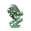
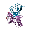
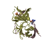
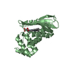
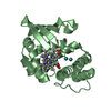
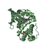
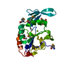
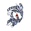
- PDB-1f7c: CRYSTAL STRUCTURE OF THE BH DOMAIN FROM GRAF, THE GTPASE REGULATO... -
+ Open data

- Basic information
| Entry | Database: PDB / ID: 1f7c | ||||||
|---|---|---|---|---|---|---|---|
| Title | CRYSTAL STRUCTURE OF THE BH DOMAIN FROM GRAF, THE GTPASE REGULATOR ASSOCIATED WITH FOCAL ADHESION KINASE | ||||||
 Components Components | RHOGAP PROTEIN RhoGAP domain RhoGAP domain | ||||||
 Keywords Keywords | SIGNALING PROTEIN / GAP / GTPase activating protein / Rho GTPase regulator / BH domain | ||||||
| Function / homology |  Function and homology information: / regulation of small GTPase mediated signal transduction / GTPase activator activity / actin cytoskeleton organization / cytoskeleton / focal adhesion / signal transduction / cytoplasm Function and homology information: / regulation of small GTPase mediated signal transduction / GTPase activator activity / actin cytoskeleton organization / cytoskeleton / focal adhesion / signal transduction / cytoplasmSimilarity search - Function | ||||||
| Biological species |  Gallus gallus (chicken) Gallus gallus (chicken) | ||||||
| Method | X-RAY DIFFRACTION / MIR, molecular replacement / Resolution: 2.4 Å | ||||||
 Authors Authors | Longenecker, K.L. / Derewenda, U. / Sheffield, P.J. / Zheng, Y. / Derewenda, Z.S. | ||||||
 Citation Citation | Journal: J.Biol.Chem. / Year: 2000 Title: Structure of the BH domain from graf and its implications for Rho GTPase recognition. Authors: Longenecker, K.L. / Zhang, B. / Derewenda, U. / Sheffield, P.J. / Dauter, Z. / Parsons, J.T. / Zheng, Y. / Derewenda, Z.S. #1: Journal: Acta Crystallogr.,Sect.D / Year: 1999Title: Expression, purification and crystallization of a BH domain from the GTPase regulatory protein associated with focal adhesion kinase Authors: Sheffield, P.J. / Derewenda, U. / Taylor, J. / Parsons, T.J. / Derewenda, Z.S. | ||||||
| History |
|
- Structure visualization
| Structure viewer | Molecule: MolmilJmol/JSmol |
|---|
- Downloads & links
-Download
| PDBx/mmCIF format | 1f7c.cif.gz | 49 KB | Display | PDBx/mmCIF format |
|---|---|---|---|---|
| PDB format | pdb1f7c.ent.gz | 34.8 KB | Display | PDB format |
| PDBx/mmJSON format | 1f7c.json.gz | Tree view | PDBx/mmJSON format | |
| Others |  Other downloads Other downloads |
-Validation report
| Arichive directory | https://data.pdbj.org/pub/pdb/validation_reports/f7/1f7cftp://data.pdbj.org/pub/pdb/validation_reports/f7/1f7c | HTTPS FTP |
|---|
-Related structure data
| Similar structure data |
|---|


-Links
 PDBj
PDBj

- Assembly
| Deposited unit | 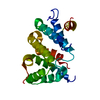
| |||||||||
|---|---|---|---|---|---|---|---|---|---|---|
| 1 |
| |||||||||
| Unit cell |
| |||||||||
| Components on special symmetry positions |
| |||||||||
| Details | monomer provides biological activity |
-Components
| #1: Protein | RhoGAP domain Mass: 26411.467 Da / Num. of mol.: 1 Fragment: GTPASE ACTIVATING PROTEIN (GAP) FOR RHO FAMILY GTPASES Source method: isolated from a genetically manipulated source Source: (gene. exp.) Gallus gallus (chicken) / Plasmid: PGEX4T1 / Production host:  Escherichia coli (E. coli) / References: UniProt: Q98935, UniProt: Q5ZMW5*PLUS Escherichia coli (E. coli) / References: UniProt: Q98935, UniProt: Q5ZMW5*PLUS |
|---|---|
| #2: Water | ChemComp-HOH / Water Mass: 18.015 Da / Num. of mol.: 47 / Source method: isolated from a natural source / Formula: H2O Mass: 18.015 Da / Num. of mol.: 47 / Source method: isolated from a natural source / Formula: H2O |
-Experimental details
-Experiment
| Experiment | Method: X-RAY DIFFRACTION / Number of used crystals: 1 |
|---|
- Sample preparation
| Crystal | Density Matthews: 2.12 Å3/Da / Density % sol: 41.88 % | ||||||||||||||||||||
|---|---|---|---|---|---|---|---|---|---|---|---|---|---|---|---|---|---|---|---|---|---|
| Crystal grow | Temperature: 294 K / Method: vapor diffusion / pH: 7 Details: PEG 6000, Na HEPES, pH 7.0, VAPOR DIFFUSION, temperature 294K | ||||||||||||||||||||
| Crystal grow | *PLUS | ||||||||||||||||||||
| Components of the solutions | *PLUS
|
-Data collection
| Diffraction | Mean temperature: 293 K |
|---|---|
| Diffraction source | Source: ROTATING ANODE / Type: ENRAF-NONIUS FR571 / Wavelength: 1.5418 |
| Detector | Type: RIGAKU RAXIS IV / Detector: IMAGE PLATE / Date: Apr 1, 1998 |
| Radiation | Protocol: SINGLE WAVELENGTH / Monochromatic (M) / Laue (L): M / Scattering type: x-ray |
| Radiation wavelength | Wavelength: 1.5418 Å / Relative weight: 1 |
| Reflection | Resolution: 2.4→20 Å / Num. all: 30884 / Num. obs: 9063 / % possible obs: 99 % / Observed criterion σ(F): 0 / Redundancy: 3.4 % / Biso Wilson estimate: 35.1 Å2 / Rmerge(I) obs: 0.077 / Net I/σ(I): 11.4 |
| Reflection shell | Resolution: 2.4→2.49 Å / Redundancy: 3 % / Rmerge(I) obs: 0.326 / Num. unique all: 866 / % possible all: 97.5 |
| Reflection | *PLUS Num. obs: 9020 / Num. measured all: 30884 |
| Reflection shell | *PLUS % possible obs: 98 % / Rmerge(I) obs: 0.33 / Mean I/σ(I) obs: 2.8 |
- Processing
| Software |
| |||||||||||||||||||||||||
|---|---|---|---|---|---|---|---|---|---|---|---|---|---|---|---|---|---|---|---|---|---|---|---|---|---|---|
| Refinement | Method to determine structure: MIR, molecular replacement / Resolution: 2.4→20 Å / σ(F): 0 / σ(I): 0 / Stereochemistry target values: Engh & Huber
| |||||||||||||||||||||||||
| Solvent computation | Solvent model: CNS 0.9 / Bsol: 42.5 Å2 | |||||||||||||||||||||||||
| Displacement parameters | Biso mean: 47 Å2 | |||||||||||||||||||||||||
| Refinement step | Cycle: LAST / Resolution: 2.4→20 Å
| |||||||||||||||||||||||||
| Refine LS restraints |
| |||||||||||||||||||||||||
| Software | *PLUS Name: CNS / Version: 0.9 / Classification: refinement | |||||||||||||||||||||||||
| Refine LS restraints | *PLUS
|
