 Movie
Movie Controller

[English] 日本語
 Yorodumi
- PDB-1ej2: Crystal structure of methanobacterium thermoautotrophicum nicotin... -
+ Open data

- Basic information
| Entry | Database: PDB / ID: 1ej2 | ||||||
|---|---|---|---|---|---|---|---|
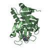
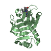
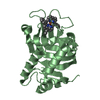
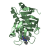
| Title | Crystal structure of methanobacterium thermoautotrophicum nicotinamide mononucleotide adenylyltransferase with bound NAD+ | ||||||
 Components Components | NICOTINAMIDE MONONUCLEOTIDE ADENYLYLTRANSFERASE | ||||||
 Keywords Keywords |  TRANSFERASE / DINUCLEOTIDE BINDING FOLD / Structural Genomics / PSI / Protein Structure Initiative / Midwest Center for Structural Genomics / MCSG / Northeast Structural Genomics Consortium / NESG TRANSFERASE / DINUCLEOTIDE BINDING FOLD / Structural Genomics / PSI / Protein Structure Initiative / Midwest Center for Structural Genomics / MCSG / Northeast Structural Genomics Consortium / NESG | ||||||
| Function / homology |  Function and homology informationnicotinamide-nucleotide adenylyltransferase / nicotinamide-nucleotide adenylyltransferase activity / NAD biosynthetic process / ATP binding / cytoplasm Function and homology informationnicotinamide-nucleotide adenylyltransferase / nicotinamide-nucleotide adenylyltransferase activity / NAD biosynthetic process / ATP binding / cytoplasmSimilarity search - Function | ||||||
| Biological species |   Methanothermobacter thermautotrophicus (archaea) Methanothermobacter thermautotrophicus (archaea) | ||||||
| Method | X-RAY DIFFRACTION / SYNCHROTRON / Resolution: 1.9 Å | ||||||
 Authors Authors | Saridakis, V. / Christendat, D. / Kimber, M.S. / Edwards, A.M. / Pai, E.F. / Midwest Center for Structural Genomics (MCSG) / Northeast Structural Genomics Consortium (NESG) | ||||||
 Citation Citation | Journal: J.Biol.Chem. / Year: 2001 Title: Insights into ligand binding and catalysis of a central step in NAD+ synthesis: structures of Methanobacterium thermoautotrophicum NMN adenylyltransferase complexes. Authors: Saridakis, V. / Christendat, D. / Kimber, M.S. / Dharamsi, A. / Edwards, A.M. / Pai, E.F. | ||||||
| History |
|
- Structure visualization
| Structure viewer | Molecule: MolmilJmol/JSmol |
|---|
- Downloads & links
-Download
| PDBx/mmCIF format | 1ej2.cif.gz | 51.1 KB | Display | PDBx/mmCIF format |
|---|---|---|---|---|
| PDB format | pdb1ej2.ent.gz | 36.3 KB | Display | PDB format |
| PDBx/mmJSON format | 1ej2.json.gz | Tree view | PDBx/mmJSON format | |
| Others |  Other downloads Other downloads |
-Validation report
| Arichive directory | https://data.pdbj.org/pub/pdb/validation_reports/ej/1ej2ftp://data.pdbj.org/pub/pdb/validation_reports/ej/1ej2 | HTTPS FTP |
|---|
-Related structure data


-Links
 PDBj
PDBj- Assembly
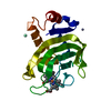
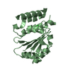
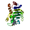
| Deposited unit | 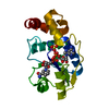
| ||||||||
|---|---|---|---|---|---|---|---|---|---|
| 1 |
| ||||||||
| 2 | x 6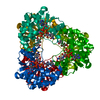
| ||||||||
| Unit cell |
|
-Components
| #1: Protein | Mass: 20596.793 Da / Num. of mol.: 1 Source method: isolated from a genetically manipulated source Source: (gene. exp.) Methanothermobacter thermautotrophicus (archaea)Plasmid: PET15B / Production host:  Escherichia coli (E. coli) Escherichia coli (E. coli)References: UniProt: O26253, nicotinamide-nucleotide adenylyltransferase |
|---|---|
| #2: Chemical | ChemComp-SO4 / Sulfate  Mass: 96.063 Da / Num. of mol.: 1 / Source method: obtained synthetically / Formula: SO4 Mass: 96.063 Da / Num. of mol.: 1 / Source method: obtained synthetically / Formula: SO4 |
| #3: Chemical | ChemComp-NA /   Mass: 22.990 Da / Num. of mol.: 1 / Source method: obtained synthetically / Formula: Na Mass: 22.990 Da / Num. of mol.: 1 / Source method: obtained synthetically / Formula: Na |
| #4: Chemical | ChemComp-NAD / Nicotinamide adenine dinucleotide  Mass: 663.425 Da / Num. of mol.: 1 / Source method: obtained synthetically / Formula: C21H27N7O14P2 / Comment: NAD*YM Mass: 663.425 Da / Num. of mol.: 1 / Source method: obtained synthetically / Formula: C21H27N7O14P2 / Comment: NAD*YM |
| #5: Water | ChemComp-HOH / Water Mass: 18.015 Da / Num. of mol.: 120 / Source method: isolated from a natural source / Formula: H2O Mass: 18.015 Da / Num. of mol.: 120 / Source method: isolated from a natural source / Formula: H2O |
-Experimental details
-Experiment
| Experiment | Method: X-RAY DIFFRACTION / Number of used crystals: 1 |
|---|
- Sample preparation
| Crystal | Density Matthews: 3.06 Å3/Da / Density % sol: 59.76 % | ||||||||||||||||||||||||
|---|---|---|---|---|---|---|---|---|---|---|---|---|---|---|---|---|---|---|---|---|---|---|---|---|---|
| Crystal grow | Method: vapor diffusion, hanging drop / pH: 7.5 Details: 1.5 M LiSO4, 0.1 M HEPES, pH 7.5, VAPOR DIFFUSION, HANGING DROP | ||||||||||||||||||||||||
| Crystal grow | *PLUS Temperature: 20 ℃ | ||||||||||||||||||||||||
| Components of the solutions | *PLUS
|
-Data collection
| Diffraction | Mean temperature: 100 K |
|---|---|
| Diffraction source | Source: SYNCHROTRON / Site: APS  / Beamline: 14-BM-C / Wavelength: 1 / Beamline: 14-BM-C / Wavelength: 1 |
| Detector | Type: ADSC QUANTUM 4 / Detector: CCD / Date: Oct 11, 1999 |
| Radiation | Protocol: SINGLE WAVELENGTH / Monochromatic (M) / Laue (L): M / Scattering type: x-ray |
| Radiation wavelength | Wavelength: 1 Å / Relative weight: 1 |
| Reflection | Resolution: 1.9→30 Å / Num. obs: 20932 / % possible obs: 100 % / Observed criterion σ(I): 521.5 / Redundancy: 11.4 % / Biso Wilson estimate: 25 Å2 / Rmerge(I) obs: 0.041 / Net I/σ(I): 37 |
| Reflection shell | Resolution: 1.9→1.97 Å / Redundancy: 6 % / Rmerge(I) obs: 0.388 / % possible all: 100 |
| Reflection | *PLUS Num. measured all: 492526 |
| Reflection shell | *PLUS % possible obs: 100 % |
- Processing
| Software |
| ||||||||||||||||||||||||||||||||||||||||||||||||||||||||||||||||||||||||||||||||
|---|---|---|---|---|---|---|---|---|---|---|---|---|---|---|---|---|---|---|---|---|---|---|---|---|---|---|---|---|---|---|---|---|---|---|---|---|---|---|---|---|---|---|---|---|---|---|---|---|---|---|---|---|---|---|---|---|---|---|---|---|---|---|---|---|---|---|---|---|---|---|---|---|---|---|---|---|---|---|---|---|---|
| Refinement | Resolution: 1.9→28.3 Å / Rfactor Rfree error: 0.005 / Data cutoff high absF: 1082435.71 / Data cutoff low absF: 0 / Isotropic thermal model: RESTRAINED / Cross valid method: THROUGHOUT / σ(F): 2 / Stereochemistry target values: CNS 0.9 / Details: SIMULATED ANNEALING
| ||||||||||||||||||||||||||||||||||||||||||||||||||||||||||||||||||||||||||||||||
| Solvent computation | Solvent model: FLAT MODEL / Bsol: 50.04 Å2 / ksol: 0.388 e/Å3 | ||||||||||||||||||||||||||||||||||||||||||||||||||||||||||||||||||||||||||||||||
| Displacement parameters | Biso mean: 36.5 Å2
| ||||||||||||||||||||||||||||||||||||||||||||||||||||||||||||||||||||||||||||||||
| Refine analyze |
| ||||||||||||||||||||||||||||||||||||||||||||||||||||||||||||||||||||||||||||||||
| Refinement step | Cycle: LAST / Resolution: 1.9→28.3 Å
| ||||||||||||||||||||||||||||||||||||||||||||||||||||||||||||||||||||||||||||||||
| Refine LS restraints |
| ||||||||||||||||||||||||||||||||||||||||||||||||||||||||||||||||||||||||||||||||
| LS refinement shell | Resolution: 1.9→2.02 Å / Rfactor Rfree error: 0.016 / Total num. of bins used: 6
| ||||||||||||||||||||||||||||||||||||||||||||||||||||||||||||||||||||||||||||||||
| Xplor file |
| ||||||||||||||||||||||||||||||||||||||||||||||||||||||||||||||||||||||||||||||||
| Software | *PLUS Name: CNS / Version: 0.5 / Classification: refinement | ||||||||||||||||||||||||||||||||||||||||||||||||||||||||||||||||||||||||||||||||
| Refine LS restraints | *PLUS
|
