 Movie
Movie Controller


+ Open data

- Basic information
| Entry | Database: PDB / ID: 1egp | ||||||
|---|---|---|---|---|---|---|---|
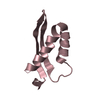
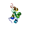
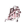
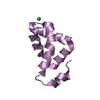
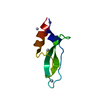
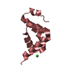
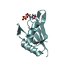
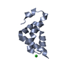
| Title | PROTEINASE INHIBITOR EGLIN C WITH HYDROLYSED REACTIVE CENTER | ||||||
 Components Components | (EGLIN-C) x 2 | ||||||
 Keywords Keywords |  PROTEINASE INHIBITOR PROTEINASE INHIBITOR | ||||||
| Function / homology | Proteinase inhibitor I13, potato inhibitor I / Proteinase inhibitor I13, potato inhibitor I superfamily / Potato inhibitor I family / Potato inhibitor I family signature. / serine-type endopeptidase inhibitor activity / response to wounding / Eglin C Function and homology information Function and homology information | ||||||
| Biological species |  Hirudo medicinalis (medicinal leech) Hirudo medicinalis (medicinal leech) | ||||||
| Method | X-RAY DIFFRACTION / Resolution: 2 Å | ||||||
 Authors Authors | Dauter, Z. / Lamzin, V. / Betzel, C. / Wilson, K.S. | ||||||
 Citation Citation | Journal: FEBS Lett. / Year: 1993 Title: Structure of the proteinase inhibitor eglin c with hydrolysed reactive centre at 2.0 A resolution. Authors: Betzel, C. / Dauter, Z. / Genov, N. / Lamzin, V. / Navaza, J. / Schnebli, H.P. / Visanji, M. / Wilson, K.S. | ||||||
| History |
|
- Structure visualization
| Structure viewer | Molecule: MolmilJmol/JSmol |
|---|
- Downloads & links
-Download
| PDBx/mmCIF format | 1egp.cif.gz | 35.5 KB | Display | PDBx/mmCIF format |
|---|---|---|---|---|
| PDB format | pdb1egp.ent.gz | 25.7 KB | Display | PDB format |
| PDBx/mmJSON format | 1egp.json.gz | Tree view | PDBx/mmJSON format | |
| Others |  Other downloads Other downloads |
-Validation report
| Arichive directory | https://data.pdbj.org/pub/pdb/validation_reports/eg/1egpftp://data.pdbj.org/pub/pdb/validation_reports/eg/1egp | HTTPS FTP |
|---|
-Related structure data
| Similar structure data |
|---|

-Links
 PDBj
PDBj- Assembly
| Deposited unit | 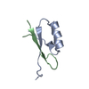
| ||||||||
|---|---|---|---|---|---|---|---|---|---|
| 1 |
| ||||||||
| Unit cell |
|
-Components
| #1: Protein/peptide | Mass: 5188.748 Da / Num. of mol.: 1 / Source method: isolated from a natural source / Details: CLEAVED BETWEEN RESIDUES 45 AND 46 / Source: (natural) Hirudo medicinalis (medicinal leech) / References: UniProt: P01051 |
|---|---|
| #2: Protein/peptide | Mass: 2928.290 Da / Num. of mol.: 1 / Source method: isolated from a natural source / Details: CLEAVED BETWEEN RESIDUES 45 AND 46 / Source: (natural) Hirudo medicinalis (medicinal leech) / References: UniProt: P01051 |
| #3: Water | ChemComp-HOH / Water Mass: 18.015 Da / Num. of mol.: 125 / Source method: isolated from a natural source / Formula: H2O Mass: 18.015 Da / Num. of mol.: 125 / Source method: isolated from a natural source / Formula: H2O |
| Compound details | SECONDARY STRUCTURE BOUNDARIES HAVE BEEN DETERMINED USING SS PROGRAM (V.S.LAMZIN, EMBL HAMBURG) AS ...SECONDARY STRUCTURE BOUNDARIES |
-Experimental details
-Experiment
| Experiment | Method: X-RAY DIFFRACTION |
|---|
- Sample preparation
| Crystal | Density Matthews: 1.9 Å3/Da / Density % sol: 35.27 % | |||||||||||||||||||||||||
|---|---|---|---|---|---|---|---|---|---|---|---|---|---|---|---|---|---|---|---|---|---|---|---|---|---|---|
| Crystal grow | *PLUS Temperature: 16 ℃ / pH: 6 / Method: vapor diffusion, hanging drop | |||||||||||||||||||||||||
| Components of the solutions | *PLUS
|
-Data collection
| Radiation | Scattering type: x-ray |
|---|---|
| Radiation wavelength | Relative weight: 1 |
| Reflection | *PLUS Highest resolution: 2 Å / Num. obs: 4131 / % possible obs: 98 % / Rmerge(I) obs: 0.06 |
- Processing
| Software |
| ||||||||||||||||||||||||||||||||||||||||||||||||||||||||||||||||||||||||||||||||||||
|---|---|---|---|---|---|---|---|---|---|---|---|---|---|---|---|---|---|---|---|---|---|---|---|---|---|---|---|---|---|---|---|---|---|---|---|---|---|---|---|---|---|---|---|---|---|---|---|---|---|---|---|---|---|---|---|---|---|---|---|---|---|---|---|---|---|---|---|---|---|---|---|---|---|---|---|---|---|---|---|---|---|---|---|---|---|
| Refinement | Resolution: 2→10 Å / σ(F): 0 /
| ||||||||||||||||||||||||||||||||||||||||||||||||||||||||||||||||||||||||||||||||||||
| Refinement step | Cycle: LAST / Resolution: 2→10 Å
| ||||||||||||||||||||||||||||||||||||||||||||||||||||||||||||||||||||||||||||||||||||
| Refine LS restraints |
|
