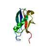
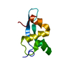
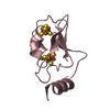
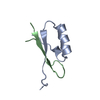
- PDB-2ljz: Structure of the C-terminal domain of HPV16 E6 oncoprotein -
+
Open data
ID or keywords:
Loading...
-
Basic information
Entry
Database: PDB / ID: 2ljz
Title
Structure of the C-terminal domain of HPV16 E6 oncoprotein
Components
Protein E6
Keywords
METAL BINDING PROTEIN
Function / homology
Function and homology information
symbiont-mediated suppression of host transcription / symbiont-mediated suppression of host apoptosis / transcription regulator activator activity / regulation of Cdc42 protein signal transduction / regulation of proteolysis / PDZ domain binding / symbiont-mediated suppression of host cytoplasmic pattern recognition receptor signaling pathway via inhibition of IRF3 activity / host cell cytoplasm / symbiont-mediated perturbation of host ubiquitin-like protein modification / symbiont-mediated suppression of host type I interferon-mediated signaling pathway ...symbiont-mediated suppression of host transcription / symbiont-mediated suppression of host apoptosis / transcription regulator activator activity / regulation of Cdc42 protein signal transduction / regulation of proteolysis / PDZ domain binding / symbiont-mediated suppression of host cytoplasmic pattern recognition receptor signaling pathway via inhibition of IRF3 activity / host cell cytoplasm / symbiont-mediated perturbation of host ubiquitin-like protein modification / symbiont-mediated suppression of host type I interferon-mediated signaling pathway / host cell nucleus / DNA-templated transcription / positive regulation of transcription by RNA polymerase II / DNA binding / zinc ion binding / identical protein binding Similarity search - Function
E6 early regulatory protein / CRO Repressor / E6 early regulatory protein / E6 superfamily / Early Protein (E6) / 2-Layer Sandwich / Alpha Beta Similarity search - Domain/homology
Mass: 65.409 Da / Num. of mol.: 1 / Source method: obtained synthetically / Formula: Zn
-
Experimental details
-
Experiment
Experiment
Method: SOLUTION NMR Details: NMR solution structure of the C-terminal zinc-binding domain of HPV16 E6
NMR experiment
Conditions-ID
Experiment-ID
Solution-ID
Type
1
1
3
2D 1H-15N HSQC
1
2
2
2D 1H-13C HSQC aliphatic
1
3
2
2D 1H-13C HSQC aromatic
1
4
2
3D HNCA
1
5
2
3D HN(CA)CB
1
6
2
3DHN(CO)CA
1
7
2
3DHBHA(CO)NH
1
8
2
3D (H)CCH-TOCSY
1
9
2
3D (H)CCH-COSY
1
10
2
3D 1H-15N NOESY
1
11
2
3D 1H-13C NOESY aliphatic
1
12
1
2D 1H-1H NOESY
1
13
1
2D 1H-1H NOESY
NMR details
Text: The structure was determined using noe and dihedral angle restraints
-
Sample preparation
Details
Solution-ID
Contents
Solvent system
1
1mME6, 90% H2O/10% D2O
90% H2O/10% D2O
2
1 mM [U-100% 13C; U-100% 15N] E6, 90% H2O/10% D2O
90% H2O/10% D2O
3
1 mM [U-100% 15N] E6, 90% H2O/10% D2O
90% H2O/10% D2O
Sample
Conc. (mg/ml)
Component
Isotopic labeling
Solution-ID
1mM
E6-1
1
1mM
E6-2
[U-100% 13C; U-100% 15N]
2
1mM
E6-3
[U-100% 15N]
3
Sample conditions
Ionic strength: 50 mM NaCl / pH: 6.8 / Pressure: ambient / Temperature: 286 K
-
NMR measurement
NMR spectrometer
Type
Manufacturer
Model
Field strength (MHz)
Spectrometer-ID
Bruker DRX
Bruker
DRX
600
1
Bruker Avance
Bruker
AVANCE
950
2
-
Processing
NMR software
Name
Version
Developer
Classification
TopSpin
2.1
BrukerBiospin
collection
NMRPipe
Delaglio, Grzesiek, Vuister, Zhu, PfeiferandBax
processing
CARA
1.8.3
KellerandWuthrich
dataanalysis
ATNOS/CANDID
Herrmann, GuntertandWuthrich
peakpicking
ATNOS/CANDID
Herrmann, GuntertandWuthrich
automaticnoeassignment
X-PLOR NIH
Schwieters, Kuszewski, TjandraandClore
structuresolution
X-PLOR NIH
Schwieters, Kuszewski, TjandraandClore
refinement
Refinement
Method: simulated annealing / Software ordinal: 1
NMR constraints
NOE constraints total: 1464 / NOE intraresidue total count: 370 / NOE long range total count: 371 / NOE medium range total count: 357 / NOE sequential total count: 366 / Protein chi angle constraints total count: 0 / Protein other angle constraints total count: 0 / Protein phi angle constraints total count: 56 / Protein psi angle constraints total count: 56
NMR representative
Selection criteria: lowest energy
NMR ensemble
Average torsion angle constraint violation: 1.15 ° Conformer selection criteria: structures with the lowest energy Conformers calculated total number: 100 / Conformers submitted total number: 20 / Maximum lower distance constraint violation: 0 Å / Maximum torsion angle constraint violation: 3.54 ° / Maximum upper distance constraint violation: 0.47 Å
NMR ensemble rms
Distance rms dev: 0.043 Å / Distance rms dev error: 0.001 Å
+
About Yorodumi
-
News
-
Feb 9, 2022. New format data for meta-information of EMDB entries
New format data for meta-information of EMDB entries
Version 3 of the EMDB header file is now the official format.
The previous official version 1.9 will be removed from the archive.
In the structure databanks used in Yorodumi, some data are registered as the other names, "COVID-19 virus" and "2019-nCoV". Here are the details of the virus and the list of structure data.
Jan 31, 2019. EMDB accession codes are about to change! (news from PDBe EMDB page)
EMDB accession codes are about to change! (news from PDBe EMDB page)
The allocation of 4 digits for EMDB accession codes will soon come to an end. Whilst these codes will remain in use, new EMDB accession codes will include an additional digit and will expand incrementally as the available range of codes is exhausted. The current 4-digit format prefixed with “EMD-” (i.e. EMD-XXXX) will advance to a 5-digit format (i.e. EMD-XXXXX), and so on. It is currently estimated that the 4-digit codes will be depleted around Spring 2019, at which point the 5-digit format will come into force.
The EM Navigator/Yorodumi systems omit the EMD- prefix.
Related info.:Q: What is EMD? / ID/Accession-code notation in Yorodumi/EM Navigator
Yorodumi is a browser for structure data from EMDB, PDB, SASBDB, etc.
This page is also the successor to EM Navigator detail page, and also detail information page/front-end page for Omokage search.
The word "yorodu" (or yorozu) is an old Japanese word meaning "ten thousand". "mi" (miru) is to see.
Related info.:EMDB / PDB / SASBDB / Comparison of 3 databanks / Yorodumi Search / Aug 31, 2016. New EM Navigator & Yorodumi / Yorodumi Papers / Jmol/JSmol / Function and homology information / Changes in new EM Navigator and Yorodumi
 Movie
Movie Controller


 Open data
 Basic information
 Components
 Keywords
 Function and homology information
 Human papillomavirus
 Authors
 Citation
 Structure visualization
 Downloads & links
 Other downloads
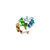


 PDBj
PDBj
 Assembly


 Mass: 65.409 Da / Num. of mol.: 1 / Source method: obtained synthetically / Formula: Zn
 HSQC
HSQC Sample preparation
 Processing