 Movie
Movie Controller

[English] 日本語
 Yorodumi
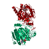
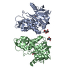
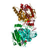
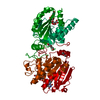
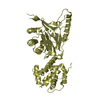
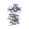
- PDB-1efh: CRYSTAL STRUCTURE OF THE HUMAN HYDROXYSTEROID SULFOTRANSFERASE IN... -
+ Open data

- Basic information
| Entry | Database: PDB / ID: 1efh | ||||||
|---|---|---|---|---|---|---|---|
| Title | CRYSTAL STRUCTURE OF THE HUMAN HYDROXYSTEROID SULFOTRANSFERASE IN THE PRESENCE OF PAP | ||||||
 Components Components | HYDROXYSTEROID SULFOTRANSFERASE | ||||||
 Keywords Keywords |  TRANSFERASE / hydroxysteroid / sulfotransferase / DHEA / A3P / PAPS / SULT2A3 TRANSFERASE / hydroxysteroid / sulfotransferase / DHEA / A3P / PAPS / SULT2A3 | ||||||
| Function / homology |  Function and homology information Function and homology informationbile-salt sulfotransferase / alcohol sulfotransferase activity / bile-salt sulfotransferase activity / alcohol sulfotransferase / steroid sulfotransferase activity / bile acid catabolic process / thyroid hormone metabolic process / 3'-phosphoadenosine 5'-phosphosulfate binding / Cytosolic sulfonation of small molecules / 3'-phosphoadenosine 5'-phosphosulfate metabolic process ...bile-salt sulfotransferase / alcohol sulfotransferase activity / bile-salt sulfotransferase activity / alcohol sulfotransferase / steroid sulfotransferase activity / bile acid catabolic process / thyroid hormone metabolic process / 3'-phosphoadenosine 5'-phosphosulfate binding / Cytosolic sulfonation of small molecules / 3'-phosphoadenosine 5'-phosphosulfate metabolic process / sulfation / ethanol catabolic process / sulfotransferase activity / Paracetamol ADME / steroid metabolic process / lipid catabolic process / cholesterol metabolic process / xenobiotic metabolic process / PPARA activates gene expression / cytosol / cytoplasmSimilarity search - Function | ||||||
| Biological species |  Homo sapiens (human) Homo sapiens (human) | ||||||
| Method | X-RAY DIFFRACTION / SYNCHROTRON / Resolution: 2.4 Å | ||||||
 Authors Authors | Pedersen, L.C. / Petrotchenko, E.V. / Negishi, M. | ||||||
 Citation Citation | Journal: FEBS Lett. / Year: 2000 Title: Crystal structure of SULT2A3, human hydroxysteroid sulfotransferase. Authors: Pedersen, L.C. / Petrotchenko, E.V. / Negishi, M. | ||||||
| History |
| ||||||
| Remark 300 | BIOMOLECULE: 1, 2 THIS ENTRY CONTAINS THE CRYSTALLOGRAPHIC ASYMMETRIC UNIT WHICH CONSISTS OF 2 ... BIOMOLECULE: 1, 2 THIS ENTRY CONTAINS THE CRYSTALLOGRAPHIC ASYMMETRIC UNIT WHICH CONSISTS OF 2 CHAIN(S). SEE REMARK 350 FOR INFORMATION ON GENERATING THE BIOLOGICAL MOLECULE(S). Despite the large surface interaction between the two molecules in the chosen asymmetric unit, the authors believe that the biological dimer is represented by the symmetry operators below. This data will be reported in the near future and referenced here at that time. |
- Structure visualization
| Structure viewer | Molecule: MolmilJmol/JSmol |
|---|
- Downloads & links
-Download
| PDBx/mmCIF format | 1efh.cif.gz | 128.4 KB | Display | PDBx/mmCIF format |
|---|---|---|---|---|
| PDB format | pdb1efh.ent.gz | 101.2 KB | Display | PDB format |
| PDBx/mmJSON format | 1efh.json.gz | Tree view | PDBx/mmJSON format | |
| Others |  Other downloads Other downloads |
-Validation report
| Arichive directory | https://data.pdbj.org/pub/pdb/validation_reports/ef/1efhftp://data.pdbj.org/pub/pdb/validation_reports/ef/1efh | HTTPS FTP |
|---|
-Related structure data
| Similar structure data |
|---|


-Links
 PDBj
PDBj
- Assembly
| Deposited unit | 
| ||||||||
|---|---|---|---|---|---|---|---|---|---|
| 1 | 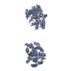
| ||||||||
| Unit cell |
|
-Components
| #1: Protein | Mass: 34545.613 Da / Num. of mol.: 2 Source method: isolated from a genetically manipulated source Source: (gene. exp.) Homo sapiens (human) / Gene: CDNA LIBRARY / Organ: LIVER / Plasmid: PET21A / Production host:  Escherichia coli (E. coli) / References: UniProt: Q06520, alcohol sulfotransferase Escherichia coli (E. coli) / References: UniProt: Q06520, alcohol sulfotransferase#2: Chemical | Adenosine 3',5'-bisphosphate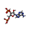  Type: RNA linking / Mass: 427.201 Da / Num. of mol.: 2 / Source method: obtained synthetically / Formula: C10H15N5O10P2 Type: RNA linking / Mass: 427.201 Da / Num. of mol.: 2 / Source method: obtained synthetically / Formula: C10H15N5O10P2#3: Water | ChemComp-HOH / | Water Mass: 18.015 Da / Num. of mol.: 185 / Source method: isolated from a natural source / Formula: H2O Mass: 18.015 Da / Num. of mol.: 185 / Source method: isolated from a natural source / Formula: H2O |
|---|
-Experimental details
-Experiment
| Experiment | Method: X-RAY DIFFRACTION / Number of used crystals: 1 |
|---|
- Sample preparation
| Crystal | Density Matthews: 3.26 Å3/Da / Density % sol: 62.33 % | ||||||||||||||||||||||||||||||
|---|---|---|---|---|---|---|---|---|---|---|---|---|---|---|---|---|---|---|---|---|---|---|---|---|---|---|---|---|---|---|---|
| Crystal grow | Temperature: 300 K / Method: vapor diffusion, hanging drop / pH: 5.75 Details: 0.8 M citrate, 80 mM cacodylate, 4 mM PAP, pH 5.75, VAPOR DIFFUSION, HANGING DROP, temperature 300K | ||||||||||||||||||||||||||||||
| Crystal grow | *PLUS pH: 7.5 / Method: unknown | ||||||||||||||||||||||||||||||
| Components of the solutions | *PLUS
|
-Data collection
| Diffraction | Mean temperature: 93 K |
|---|---|
| Diffraction source | Source: SYNCHROTRON / Site: NSLS  / Beamline: X9B / Wavelength: 0.9786 / Beamline: X9B / Wavelength: 0.9786 |
| Detector | Type: ADSC QUANTUM 4 / Detector: CCD / Date: Aug 27, 1999 |
| Radiation | Protocol: SINGLE WAVELENGTH / Monochromatic (M) / Laue (L): M / Scattering type: x-ray |
| Radiation wavelength | Wavelength: 0.9786 Å / Relative weight: 1 |
| Reflection | Resolution: 2.4→25 Å / Num. obs: 113997 / % possible obs: 94.4 % / Observed criterion σ(I): -3 / Redundancy: 3.34 % / Biso Wilson estimate: 29.6 Å2 / Rmerge(I) obs: 0.05 / Net I/σ(I): 15.3 |
| Reflection shell | Resolution: 2.4→2.49 Å / Redundancy: 2.4 % / Rmerge(I) obs: 0.146 / Num. unique all: 3087 / % possible all: 86.9 |
| Reflection | *PLUS Num. obs: 34110 / Num. measured all: 113997 / Rmerge(I) obs: 0.05 |
| Reflection shell | *PLUS % possible obs: 86.9 % / Mean I/σ(I) obs: 6.7 |
- Processing
| Software |
| ||||||||||||||||||||
|---|---|---|---|---|---|---|---|---|---|---|---|---|---|---|---|---|---|---|---|---|---|
| Refinement | Resolution: 2.4→20 Å / Rfactor Rfree error: 0.006 / Data cutoff high absF: 585066.79 / Data cutoff high rms absF: 585066.79 / Data cutoff low absF: 0 / Isotropic thermal model: RESTRAINED / Cross valid method: THROUGHOUT / σ(F): 0
| ||||||||||||||||||||
| Solvent computation | Solvent model: FLAT MODEL / Bsol: 29.18 Å2 / ksol: 0.316 e/Å3 | ||||||||||||||||||||
| Displacement parameters | Biso mean: 36.9 Å2
| ||||||||||||||||||||
| Refine analyze |
| ||||||||||||||||||||
| Refinement step | Cycle: LAST / Resolution: 2.4→20 Å
| ||||||||||||||||||||
| Refine LS restraints |
| ||||||||||||||||||||
| LS refinement shell | Resolution: 2.4→2.55 Å / Rfactor Rfree error: 0.019 / Total num. of bins used: 6
| ||||||||||||||||||||
| Xplor file |
| ||||||||||||||||||||
| Software | *PLUS Name: CNS / Version: 0.5 / Classification: refinement | ||||||||||||||||||||
| Refinement | *PLUS σ(F): 0 / % reflection Rfree: 4.9 % | ||||||||||||||||||||
| Solvent computation | *PLUS | ||||||||||||||||||||
| Displacement parameters | *PLUS Biso mean: 36.9 Å2 | ||||||||||||||||||||
| Refine LS restraints | *PLUS
| ||||||||||||||||||||
| LS refinement shell | *PLUS Rfactor Rfree: 0.303 / % reflection Rfree: 4.9 % / Rfactor Rwork: 0.248 |
