 Movie
Movie Controller


+ Open data

- Basic information
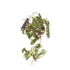
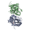
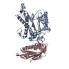
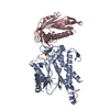
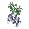
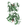
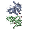
| Entry | Database: PDB / ID: 4qep | ||||||
|---|---|---|---|---|---|---|---|
| Title | crystal structure of KRYPTONITE in complex with mCHG DNA and SAH | ||||||
 Components Components |
| ||||||
 Keywords Keywords | transcription/DNA / SRA / SET / Histone methylation / methylated DNA / Methylation / transcription-DNA complex | ||||||
| Function / homology |  Function and homology information Function and homology informationmethyl-CpNpG binding / methyl-CpNpN binding / peptidyl-lysine methylation / [histone H3]-lysine9 N-methyltransferase / histone H3K9 methyltransferase activity / histone H3K9 monomethyltransferase activity / double-stranded methylated DNA binding / methyl-CpG binding / histone methyltransferase activity / negative regulation of gene expression via chromosomal CpG island methylation ...methyl-CpNpG binding / methyl-CpNpN binding / peptidyl-lysine methylation / [histone H3]-lysine9 N-methyltransferase / histone H3K9 methyltransferase activity / histone H3K9 monomethyltransferase activity / double-stranded methylated DNA binding / methyl-CpG binding / histone methyltransferase activity / negative regulation of gene expression via chromosomal CpG island methylation / chromosome, centromeric region / Transferases; Transferring one-carbon groups; Methyltransferases / double-stranded DNA binding / zinc ion binding / nucleus Similarity search - Function | ||||||
| Biological species |  | ||||||
| Method |  X-RAY DIFFRACTION / SYNCHROTRON / MOLECULAR REPLACEMENT / Resolution: 3.1 Å X-RAY DIFFRACTION / SYNCHROTRON / MOLECULAR REPLACEMENT / Resolution: 3.1 Å | ||||||
 Authors Authors | Du, J. / Li, S. / Patel, D.J. | ||||||
 Citation Citation | Journal: Mol.Cell / Year: 2014 Title: Mechanism of DNA Methylation-Directed Histone Methylation by KRYPTONITE. Authors: Du, J. / Johnson, L.M. / Groth, M. / Feng, S. / Hale, C.J. / Li, S. / Vashisht, A.A. / Gallego-Bartolome, J. / Wohlschlegel, J.A. / Patel, D.J. / Jacobsen, S.E. | ||||||
| History |
|
- Structure visualization
| Structure viewer | Molecule: MolmilJmol/JSmol |
|---|
- Downloads & links
-Download
| PDBx/mmCIF format | 4qep.cif.gz | 231.5 KB | Display | PDBx/mmCIF format |
|---|---|---|---|---|
| PDB format | pdb4qep.ent.gz | 180.8 KB | Display | PDB format |
| PDBx/mmJSON format | 4qep.json.gz | Tree view | PDBx/mmJSON format | |
| Others |  Other downloads Other downloads |
-Validation report
| Arichive directory | https://data.pdbj.org/pub/pdb/validation_reports/qe/4qepftp://data.pdbj.org/pub/pdb/validation_reports/qe/4qep | HTTPS FTP |
|---|
-Related structure data
| Related structure data |  4qenSC  4qeoC S: Starting model for refinement C: citing same article ( |
|---|---|
| Similar structure data |


-Links
 PDBj
PDBj


- Assembly
| Deposited unit | 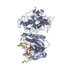
| ||||||||
|---|---|---|---|---|---|---|---|---|---|
| 1 |
| ||||||||
| Unit cell |
| ||||||||
| Details | The biological assembly is the asymmetric unit. |
-Components
| #1: Protein | Mass: 59686.797 Da / Num. of mol.: 1 / Fragment: functional fragment Source method: isolated from a genetically manipulated source Source: (gene. exp.)  References: UniProt: Q8GZB6, histone-lysine N-methyltransferase |
|---|---|
| #2: DNA chain | Mass: 4623.037 Da / Num. of mol.: 1 / Source method: obtained synthetically |
| #3: DNA chain | Mass: 4568.985 Da / Num. of mol.: 1 / Source method: obtained synthetically |
| #4: Chemical | ChemComp-SAH /   Type: L-peptide linking / Mass: 384.411 Da / Num. of mol.: 1 / Source method: obtained synthetically / Formula: C14H20N6O5S Type: L-peptide linking / Mass: 384.411 Da / Num. of mol.: 1 / Source method: obtained synthetically / Formula: C14H20N6O5S |
| #5: Chemical | ChemComp-ZN /   Mass: 65.409 Da / Num. of mol.: 4 / Source method: obtained synthetically / Formula: Zn Mass: 65.409 Da / Num. of mol.: 4 / Source method: obtained synthetically / Formula: Zn |
-Experimental details
-Experiment
| Experiment | Method: X-RAY DIFFRACTION / Number of used crystals: 1 |
|---|
- Sample preparation
| Crystal | Density Matthews: 2.3 Å3/Da / Density % sol: 46.41 % |
|---|---|
| Crystal grow | Temperature: 293 K / Method: vapor diffusion, hanging drop / pH: 6 Details: 30% PEG200, 5% PEG3000, and 0.1 M MES, pH 6.0, VAPOR DIFFUSION, HANGING DROP, temperature 293K |
-Data collection
| Diffraction | Mean temperature: 100 K |
|---|---|
| Diffraction source | Source: SYNCHROTRON / Site: APS  / Beamline: 24-ID-E / Wavelength: 0.9793 Å / Beamline: 24-ID-E / Wavelength: 0.9793 Å |
| Detector | Type: ADSC QUANTUM 315r / Detector: CCD / Date: Mar 4, 2013 |
| Radiation | Monochromator: double crystal monochrometer / Protocol: SINGLE WAVELENGTH / Monochromatic (M) / Laue (L): M / Scattering type: x-ray |
| Radiation wavelength | Wavelength: 0.9793 Å / Relative weight: 1 |
| Reflection | Resolution: 3.1→50 Å / Num. all: 12077 / Num. obs: 10851 / % possible obs: 89.6 % / Observed criterion σ(F): 0 / Observed criterion σ(I): 0 / Redundancy: 3.6 % / Rmerge(I) obs: 0.155 / Rsym value: 0.155 / Net I/σ(I): 9.5 |
| Reflection shell | Resolution: 3.1→3.21 Å / Redundancy: 3.5 % / Rmerge(I) obs: 0.615 / Mean I/σ(I) obs: 2 / Rsym value: 0.615 / % possible all: 92.8 |
- Processing
| Software |
| |||||||||||||||||||||||||||||||||||||||||||||||||||||||||||||||||||||||||||||||||||||||||||||||||||||||||||||||||||||||||||||||||||||||||||||||||||||||||||||||||||||||||||||||
|---|---|---|---|---|---|---|---|---|---|---|---|---|---|---|---|---|---|---|---|---|---|---|---|---|---|---|---|---|---|---|---|---|---|---|---|---|---|---|---|---|---|---|---|---|---|---|---|---|---|---|---|---|---|---|---|---|---|---|---|---|---|---|---|---|---|---|---|---|---|---|---|---|---|---|---|---|---|---|---|---|---|---|---|---|---|---|---|---|---|---|---|---|---|---|---|---|---|---|---|---|---|---|---|---|---|---|---|---|---|---|---|---|---|---|---|---|---|---|---|---|---|---|---|---|---|---|---|---|---|---|---|---|---|---|---|---|---|---|---|---|---|---|---|---|---|---|---|---|---|---|---|---|---|---|---|---|---|---|---|---|---|---|---|---|---|---|---|---|---|---|---|---|---|---|---|---|
| Refinement | Method to determine structure: MOLECULAR REPLACEMENT Starting model: pdb entry 4QEN Resolution: 3.1→49.627 Å / SU ML: 0.44 / σ(F): 1.37 / Phase error: 27.01 / Stereochemistry target values: ML
| |||||||||||||||||||||||||||||||||||||||||||||||||||||||||||||||||||||||||||||||||||||||||||||||||||||||||||||||||||||||||||||||||||||||||||||||||||||||||||||||||||||||||||||||
| Solvent computation | Shrinkage radii: 0.9 Å / VDW probe radii: 1.11 Å / Solvent model: FLAT BULK SOLVENT MODEL | |||||||||||||||||||||||||||||||||||||||||||||||||||||||||||||||||||||||||||||||||||||||||||||||||||||||||||||||||||||||||||||||||||||||||||||||||||||||||||||||||||||||||||||||
| Refinement step | Cycle: LAST / Resolution: 3.1→49.627 Å
| |||||||||||||||||||||||||||||||||||||||||||||||||||||||||||||||||||||||||||||||||||||||||||||||||||||||||||||||||||||||||||||||||||||||||||||||||||||||||||||||||||||||||||||||
| Refine LS restraints |
| |||||||||||||||||||||||||||||||||||||||||||||||||||||||||||||||||||||||||||||||||||||||||||||||||||||||||||||||||||||||||||||||||||||||||||||||||||||||||||||||||||||||||||||||
| LS refinement shell |
| |||||||||||||||||||||||||||||||||||||||||||||||||||||||||||||||||||||||||||||||||||||||||||||||||||||||||||||||||||||||||||||||||||||||||||||||||||||||||||||||||||||||||||||||
| Refinement TLS params. | Method: refined / Refine-ID: X-RAY DIFFRACTION
| |||||||||||||||||||||||||||||||||||||||||||||||||||||||||||||||||||||||||||||||||||||||||||||||||||||||||||||||||||||||||||||||||||||||||||||||||||||||||||||||||||||||||||||||
| Refinement TLS group |
|
