 Movie
Movie Controller

[English] 日本語
 Yorodumi
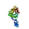
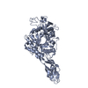
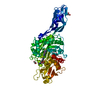
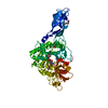
- PDB-1edq: CRYSTAL STRUCTURE OF CHITINASE A FROM S. MARCESCENS AT 1.55 ANGSTROMS -
+ Open data

- Basic information
| Entry | Database: PDB / ID: 1edq | ||||||
|---|---|---|---|---|---|---|---|
| Title | CRYSTAL STRUCTURE OF CHITINASE A FROM S. MARCESCENS AT 1.55 ANGSTROMS | ||||||
 Components Components | CHITINASE A | ||||||
 Keywords Keywords | HYDROLASE / BETA-ALPHA (TIM) BARREL | ||||||
| Function / homology |  Function and homology information Function and homology informationendochitinase activity / chitinase / chitin catabolic process / chitin binding / polysaccharide catabolic process Similarity search - Function | ||||||
| Biological species |  Serratia marcescens (bacteria) Serratia marcescens (bacteria) | ||||||
| Method |  X-RAY DIFFRACTION / SYNCHROTRON / MOLECULAR REPLACEMENT / Resolution: 1.55 Å X-RAY DIFFRACTION / SYNCHROTRON / MOLECULAR REPLACEMENT / Resolution: 1.55 Å | ||||||
 Authors Authors | Papanikolau, Y. / Petratos, K. | ||||||
 Citation Citation | Journal: Acta Crystallogr.,Sect.D / Year: 2003 Title: De novo purification scheme and crystallization conditions yield high-resolution structures of chitinase A and its complex with the inhibitor allosamidin. Authors: Papanikolau, Y. / Tavlas, G. / Vorgias, C.E. / Petratos, K. #1: Journal: Structure / Year: 1994Title: Crystal structure of a bacterial chitinase at 2.3 Angstrom resolution Authors: Perrakis, A. / Tews, I. / Dauter, Z. / Oppenheim, A.B. / Chet, I. / Wilson, K.S. / Vorgias, C.E. #2: Journal: Biochemistry / Year: 2001Title: HIGH RESOLUTION STRUCTURAL ANALYSES OF MUTANT CHITINASE A COMPLEXES WITH SUBSTRATES PROVIDE NEW INSIGHT INTO THE MECHANISM OF CATALYSIS Authors: PAPANIKOLAU, Y. / PRAG, G. / TAVLAS, G. / VORGIAS, C.E. / OPPENHEIM, A.B. / PETRATOS, K. | ||||||
| History |
|
- Structure visualization
| Structure viewer | Molecule: MolmilJmol/JSmol |
|---|
- Downloads & links
-Download
| PDBx/mmCIF format | 1edq.cif.gz | 140.2 KB | Display | PDBx/mmCIF format |
|---|---|---|---|---|
| PDB format | pdb1edq.ent.gz | 105.1 KB | Display | PDB format |
| PDBx/mmJSON format | 1edq.json.gz | Tree view | PDBx/mmJSON format | |
| Others |  Other downloads Other downloads |
-Validation report
| Arichive directory | https://data.pdbj.org/pub/pdb/validation_reports/ed/1edqftp://data.pdbj.org/pub/pdb/validation_reports/ed/1edq | HTTPS FTP |
|---|
-Related structure data
| Related structure data |  1ffqC 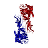 1ctnS S: Starting model for refinement C: citing same article ( |
|---|---|
| Similar structure data |


-Links
 PDBj
PDBj- Assembly
| Deposited unit | 
| ||||||||
|---|---|---|---|---|---|---|---|---|---|
| 1 |
| ||||||||
| Unit cell |
|
-Components
| #1: Protein | Mass: 58639.590 Da / Num. of mol.: 1 Source method: isolated from a genetically manipulated source Source: (gene. exp.) Serratia marcescens (bacteria) / Production host: |
|---|---|
| #2: Water | ChemComp-HOH /  Mass: 18.015 Da / Num. of mol.: 909 / Source method: isolated from a natural source / Formula: H2O Mass: 18.015 Da / Num. of mol.: 909 / Source method: isolated from a natural source / Formula: H2O |
| Has protein modification | Y |
-Experimental details
-Experiment
| Experiment | Method: X-RAY DIFFRACTION / Number of used crystals: 1 |
|---|
- Sample preparation
| Crystal | Density Matthews: 3.3 Å3/Da / Density % sol: 63 % | ||||||||||||||||||||||||||||||||||||||||||
|---|---|---|---|---|---|---|---|---|---|---|---|---|---|---|---|---|---|---|---|---|---|---|---|---|---|---|---|---|---|---|---|---|---|---|---|---|---|---|---|---|---|---|---|
| Crystal grow | Temperature: 293 K / Method: vapor diffusion, hanging drop / pH: 7.2 Details: 0.75 M Citrate-Na pH 7.2, 20% (v/v) methanol, VAPOR DIFFUSION, HANGING DROP, temperature 293K | ||||||||||||||||||||||||||||||||||||||||||
| Crystal grow | *PLUS Temperature: 291 K / pH: 7.5 | ||||||||||||||||||||||||||||||||||||||||||
| Components of the solutions | *PLUS
|
-Data collection
| Diffraction | Mean temperature: 100 K |
|---|---|
| Diffraction source | Source: SYNCHROTRON / Site: EMBL/DESY, HAMBURG  / Beamline: X11 / Wavelength: 0.9116 / Beamline: X11 / Wavelength: 0.9116 |
| Detector | Type: MARRESEARCH / Detector: IMAGE PLATE / Date: Apr 4, 1999 |
| Radiation | Protocol: SINGLE WAVELENGTH / Monochromatic (M) / Laue (L): M / Scattering type: x-ray |
| Radiation wavelength | Wavelength: 0.9116 Å / Relative weight: 1 |
| Reflection | Resolution: 1.55→10 Å / Num. all: 112857 / Num. obs: 112857 / % possible obs: 99.8 % / Observed criterion σ(F): 0 / Observed criterion σ(I): 0 / Redundancy: 5 % / Biso Wilson estimate: 19.6 Å2 / Rmerge(I) obs: 0.032 / Net I/σ(I): 27.8 |
| Reflection shell | Resolution: 1.55→1.61 Å / Redundancy: 4.3 % / Rmerge(I) obs: 0.216 / Mean I/σ(I) obs: 6.8 / Num. unique all: 11176 / % possible all: 100 |
| Reflection | *PLUS |
| Reflection shell | *PLUS % possible obs: 100 % |
- Processing
| Software |
| ||||||||||||||||||||||||||||||||||||||||||||||||||||||||||||||||||||||||||||||||
|---|---|---|---|---|---|---|---|---|---|---|---|---|---|---|---|---|---|---|---|---|---|---|---|---|---|---|---|---|---|---|---|---|---|---|---|---|---|---|---|---|---|---|---|---|---|---|---|---|---|---|---|---|---|---|---|---|---|---|---|---|---|---|---|---|---|---|---|---|---|---|---|---|---|---|---|---|---|---|---|---|---|
| Refinement | Method to determine structure: MOLECULAR REPLACEMENT Starting model: 1CTN Resolution: 1.55→10 Å / SU B: 1.328 / SU ML: 0.049 / σ(F): 0 / σ(I): 0 / ESU R: 0.072 / ESU R Free: 0.074 / Stereochemistry target values: Engh & Huber
| ||||||||||||||||||||||||||||||||||||||||||||||||||||||||||||||||||||||||||||||||
| Displacement parameters | Biso mean: 24.2 Å2 | ||||||||||||||||||||||||||||||||||||||||||||||||||||||||||||||||||||||||||||||||
| Refinement step | Cycle: LAST / Resolution: 1.55→10 Å
| ||||||||||||||||||||||||||||||||||||||||||||||||||||||||||||||||||||||||||||||||
| Refine LS restraints |
| ||||||||||||||||||||||||||||||||||||||||||||||||||||||||||||||||||||||||||||||||
| Software | *PLUS Name: REFMAC / Classification: refinement | ||||||||||||||||||||||||||||||||||||||||||||||||||||||||||||||||||||||||||||||||
| Refinement | *PLUS Lowest resolution: 10 Å / σ(F): 0 / % reflection Rfree: 5 % / Rfactor obs: 0.187 | ||||||||||||||||||||||||||||||||||||||||||||||||||||||||||||||||||||||||||||||||
| Solvent computation | *PLUS | ||||||||||||||||||||||||||||||||||||||||||||||||||||||||||||||||||||||||||||||||
| Displacement parameters | *PLUS | ||||||||||||||||||||||||||||||||||||||||||||||||||||||||||||||||||||||||||||||||
| Refine LS restraints | *PLUS
|
