 Movie
Movie Controller


+ Open data

- Basic information
| Entry | Database: PDB / ID: 1k9t | |||||||||
|---|---|---|---|---|---|---|---|---|---|---|
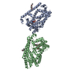
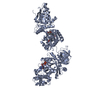
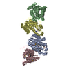
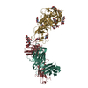
| Title | Chitinase a complexed with tetra-N-acetylchitotriose | |||||||||
 Components Components | CHITINASE A | |||||||||
 Keywords Keywords | HYDROLASE / GLYCOSYL HYDROLASE FAMILY 18 / CHITINASE / CHITINOLYSIS / A/B-(TIM)-BARREL | |||||||||
| Function / homology |  Function and homology information Function and homology informationendochitinase activity / chitinase / chitin catabolic process / chitin binding / polysaccharide catabolic process Similarity search - Function | |||||||||
| Biological species |  SERRATIA MARCESCENS (bacteria) SERRATIA MARCESCENS (bacteria) | |||||||||
| Method |  X-RAY DIFFRACTION / SYNCHROTRON / MOLECULAR REPLACEMENT / Resolution: 1.8 Å X-RAY DIFFRACTION / SYNCHROTRON / MOLECULAR REPLACEMENT / Resolution: 1.8 Å | |||||||||
 Authors Authors | Prag, G. / Tucker, P.A. / Oppenheim, A.B. | |||||||||
 Citation Citation | Journal: To be Published Title: Complex Structures of Chitinase A Mutant with Oligonag Provide Insight Into the Enzymatic Mechanism Authors: Prag, G. / Tucker, P.A. / Oppenheim, A.B. #1: Journal: Chitin Enzymol. / Year: 2001Title: Conservation of Structural Elements and Catalytic Mechanism in the Chitinolytic Enzymes from Serratia Marcescens Authors: Prag, G. / Vorgias, C.E. / Oppenheim, A.B. #2: Journal: Biochemistry / Year: 2001Title: High Resolution Structural Analyses of Mutant Chitinase a Complexes with Substrates Provide New Insight Into the Mechanism of Catalysis Authors: Papanikolau, Y. / Prag, G. / Tavlas, G. / Vorgias, C.E. / Oppenheim, A.B. / Petratos, K. #3: Journal: J.Am.Chem.Soc. / Year: 1997Title: Substrate-Assisted Catalysis Unifies Two Families of Chitinolytic Enzymes Authors: Tews, I. / Terwisscha Van Scheltinga, A.C. / Perrakis, A. / Wilson, K.S. / Dijkstra, B.W. #4: Journal: Structure / Year: 1994Title: Crystal Structure of a Bacterial Chitinase at 2.3 Angstrom Resolution Authors: Perrakis, A. / Tews, I. / Dauter, Z. / Oppenheim, A.B. / Chet, I. / Wilson, K.S. / Vorgias, C.E. | |||||||||
| History |
|
- Structure visualization
| Structure viewer | Molecule: MolmilJmol/JSmol |
|---|
- Downloads & links
-Download
| PDBx/mmCIF format | 1k9t.cif.gz | 140.1 KB | Display | PDBx/mmCIF format |
|---|---|---|---|---|
| PDB format | pdb1k9t.ent.gz | 104.3 KB | Display | PDB format |
| PDBx/mmJSON format | 1k9t.json.gz | Tree view | PDBx/mmJSON format | |
| Others |  Other downloads Other downloads |
-Validation report
| Arichive directory | https://data.pdbj.org/pub/pdb/validation_reports/k9/1k9tftp://data.pdbj.org/pub/pdb/validation_reports/k9/1k9t | HTTPS FTP |
|---|
-Related structure data
| Related structure data |  1edqS S: Starting model for refinement |
|---|---|
| Similar structure data |


-Links
 PDBj
PDBj- Assembly
| Deposited unit | 
| ||||||||
|---|---|---|---|---|---|---|---|---|---|
| 1 | 
| ||||||||
| Unit cell |
|
-Components
| #1: Protein | Mass: 58595.582 Da / Num. of mol.: 1 / Mutation: D391A Source method: isolated from a genetically manipulated source Source: (gene. exp.) SERRATIA MARCESCENS (bacteria) / Gene: CHIA / Plasmid: PKK233-3 / Production host: References: UniProt: O83008, UniProt: P07254*PLUS, chitinase |
|---|---|
| #2: Polysaccharide | 2-acetamido-2-deoxy-beta-D-glucopyranose-(1-4)-2-acetamido-2-deoxy-beta-D-glucopyranose-(1-4)-2- ...2-acetamido-2-deoxy-beta-D-glucopyranose-(1-4)-2-acetamido-2-deoxy-beta-D-glucopyranose-(1-4)-2-acetamido-2-deoxy-beta-D-glucopyranose-(1-4)-2-acetamido-2-deoxy-beta-D-glucopyranose Source method: isolated from a genetically manipulated source |
| #3: Water | ChemComp-HOH /  Mass: 18.015 Da / Num. of mol.: 761 / Source method: isolated from a natural source / Formula: H2O Mass: 18.015 Da / Num. of mol.: 761 / Source method: isolated from a natural source / Formula: H2O |
| Has protein modification | Y |
| Sequence details | THE DEPOSITORS FOUND THAT RESIDUE 475 IS GLY AND THAT IT IS VARIED FROM THE SEQUENCE IN GENBANK AT ...THE DEPOSITORS |
-Experimental details
-Experiment
| Experiment | Method: X-RAY DIFFRACTION / Number of used crystals: 1 |
|---|
- Sample preparation
| Crystal | Density Matthews: 3.35 Å3/Da / Density % sol: 63 % |
|---|---|
| Crystal grow | Temperature: 291 K / Method: vapor diffusion, hanging drop / pH: 7.2 Details: 0.75M CITRATE-NA AND 20%(V/V) METHANOL, pH 7.20, VAPOR DIFFUSION, HANGING DROP, temperature 291K |
-Data collection
| Diffraction | Mean temperature: 100 K |
|---|---|
| Diffraction source | Source: SYNCHROTRON / Site: EMBL/DESY, HAMBURG  / Beamline: X13 / Wavelength: 0.81 / Beamline: X13 / Wavelength: 0.81 |
| Detector | Type: MAR CCD 165 mm / Detector: CCD / Date: Sep 28, 2000 |
| Radiation | Monochromator: SI 111 / Protocol: SINGLE WAVELENGTH / Monochromatic (M) / Laue (L): M / Scattering type: x-ray |
| Radiation wavelength | Wavelength: 0.81 Å / Relative weight: 1 |
| Reflection | Resolution: 1.8→20 Å / Num. all: 73268 / Num. obs: 73268 / % possible obs: 100 % / Observed criterion σ(I): 0 / Redundancy: 4.3 % / Rmerge(I) obs: 0.0391 / Net I/σ(I): 18.22 |
| Reflection shell | Resolution: 1.8→1.85 Å / % possible all: 100 |
- Processing
| Software |
| ||||||||||||||||||||||||||||||||||||||||||||||||||||||||
|---|---|---|---|---|---|---|---|---|---|---|---|---|---|---|---|---|---|---|---|---|---|---|---|---|---|---|---|---|---|---|---|---|---|---|---|---|---|---|---|---|---|---|---|---|---|---|---|---|---|---|---|---|---|---|---|---|---|
| Refinement | Method to determine structure: MOLECULAR REPLACEMENT Starting model: PDB ENTRY 1EDQ Resolution: 1.8→20 Å / Cross valid method: THROUGHOUT / σ(F): 0 / Stereochemistry target values: ENGH & HUBER
| ||||||||||||||||||||||||||||||||||||||||||||||||||||||||
| Displacement parameters | Biso mean: 23.736 Å2 | ||||||||||||||||||||||||||||||||||||||||||||||||||||||||
| Refinement step | Cycle: LAST / Resolution: 1.8→20 Å
| ||||||||||||||||||||||||||||||||||||||||||||||||||||||||
| Refine LS restraints |
|
