 Movie
Movie Controller

[English] 日本語
 Yorodumi
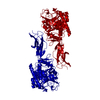
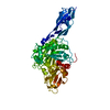
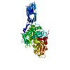
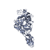
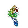
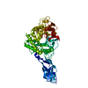
- PDB-2wly: Chitinase A from Serratia marcescens ATCC990 in complex with Chit... -
+ Open data

- Basic information
| Entry | Database: PDB / ID: 2wly | ||||||
|---|---|---|---|---|---|---|---|
| Title | Chitinase A from Serratia marcescens ATCC990 in complex with Chitotrio-thiazoline. | ||||||
 Components Components | Chitinase | ||||||
 Keywords Keywords | HYDROLASE / THIAZOLINES / CHITINASE A / GLYCOSIDASE / CHITIN HYDROLYSIS / CHITIN DEGRADATION / POLYSACCHARIDE DEGRADATION / FAMILY 18 CHITINASES / CARBOHYDRATE METABOLISM | ||||||
| Function / homology |  Function and homology information Function and homology informationendochitinase activity / chitinase / chitin catabolic process / chitin binding / polysaccharide catabolic process Similarity search - Function | ||||||
| Biological species |  Serratia marcescens (bacteria) Serratia marcescens (bacteria) | ||||||
| Method |  X-RAY DIFFRACTION / SYNCHROTRON / MOLECULAR REPLACEMENT / Resolution: 2.4 Å X-RAY DIFFRACTION / SYNCHROTRON / MOLECULAR REPLACEMENT / Resolution: 2.4 Å | ||||||
 Authors Authors | Taylor, E.J. / Dennis, R.J. / Macdonald, J.M. / Tarling, C.A. / Knapp, S. / Withers, S.G. / Davies, G.J. | ||||||
 Citation Citation | Journal: Angew. Chem. Int. Ed. Engl. / Year: 2010 Title: Chitinase inhibition by chitobiose and chitotriose thiazolines. Authors: Macdonald, J.M. / Tarling, C.A. / Taylor, E.J. / Dennis, R.J. / Myers, D.S. / Knapp, S. / Davies, G.J. / Withers, S.G. | ||||||
| History |
| ||||||
| Remark 700 | SHEET DETERMINATION METHOD: DSSP THE SHEETS PRESENTED AS "AD" IN EACH CHAIN ON SHEET RECORDS BELOW ... SHEET DETERMINATION METHOD: DSSP THE SHEETS PRESENTED AS "AD" IN EACH CHAIN ON SHEET RECORDS BELOW IS ACTUALLY AN 8-STRANDED BARREL THIS IS REPRESENTED BY A 9-STRANDED SHEET IN WHICH THE FIRST AND LAST STRANDS ARE IDENTICAL. THE SHEET STRUCTURE OF THIS MOLECULE IS BIFURCATED. IN ORDER TO REPRESENT THIS FEATURE IN THE SHEET RECORDS BELOW, TWO SHEETS ARE DEFINED. |
- Structure visualization
| Structure viewer | Molecule: MolmilJmol/JSmol |
|---|
- Downloads & links
-Download
| PDBx/mmCIF format | 2wly.cif.gz | 129.7 KB | Display | PDBx/mmCIF format |
|---|---|---|---|---|
| PDB format | pdb2wly.ent.gz | 97.8 KB | Display | PDB format |
| PDBx/mmJSON format | 2wly.json.gz | Tree view | PDBx/mmJSON format | |
| Others |  Other downloads Other downloads |
-Validation report
| Arichive directory | https://data.pdbj.org/pub/pdb/validation_reports/wl/2wlyftp://data.pdbj.org/pub/pdb/validation_reports/wl/2wly | HTTPS FTP |
|---|
-Related structure data
| Related structure data |  2wk2C  2wlzC  2wm0C  1edqS C: citing same article ( S: Starting model for refinement |
|---|---|
| Similar structure data |


-Links
 PDBj
PDBj
- Assembly
| Deposited unit | 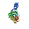
| ||||||||
|---|---|---|---|---|---|---|---|---|---|
| 1 |
| ||||||||
| Unit cell |
|
-Components
-Protein / Sugars , 2 types, 2 molecules A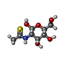
| #1: Protein | Mass: 59756.781 Da / Num. of mol.: 1 / Fragment: RESIDUES 2-520 Source method: isolated from a genetically manipulated source Source: (gene. exp.) Serratia marcescens (bacteria) / Production host: |
|---|---|
| #4: Sugar | ChemComp-SN5 /  Type: D-saccharide, beta linking / Mass: 237.273 Da / Num. of mol.: 1 / Source method: obtained synthetically / Formula: C8H15NO5S Type: D-saccharide, beta linking / Mass: 237.273 Da / Num. of mol.: 1 / Source method: obtained synthetically / Formula: C8H15NO5S |
-Non-polymers , 4 types, 335 molecules 


| #2: Chemical | ChemComp-DIO /  Mass: 88.105 Da / Num. of mol.: 5 / Source method: obtained synthetically / Formula: C4H8O2 Mass: 88.105 Da / Num. of mol.: 5 / Source method: obtained synthetically / Formula: C4H8O2#3: Chemical |  Mass: 106.120 Da / Num. of mol.: 3 / Source method: obtained synthetically / Formula: C4H10O3 Mass: 106.120 Da / Num. of mol.: 3 / Source method: obtained synthetically / Formula: C4H10O3#5: Chemical | ChemComp-NGT / |  Mass: 219.258 Da / Num. of mol.: 1 / Source method: obtained synthetically / Formula: C8H13NO4S Mass: 219.258 Da / Num. of mol.: 1 / Source method: obtained synthetically / Formula: C8H13NO4S#6: Water | ChemComp-HOH / | Mass: 18.015 Da / Num. of mol.: 326 / Source method: isolated from a natural source / Formula: H2O |
|---|
-Details
| Has protein modification | Y |
|---|---|
| Sequence details | THERE ARE KNOWN DISCREPANCIES IN THE SEQUENCE DEPENDING ON WHICH STRAIN OF SERRATIA MARCESCENS THE ...THERE ARE KNOWN DISCREPANC |
-Experimental details
-Experiment
| Experiment | Method: X-RAY DIFFRACTION / Number of used crystals: 1 |
|---|
- Sample preparation
| Crystal | Density Matthews: 3.38 Å3/Da / Density % sol: 63.62 % / Description: NONE |
|---|---|
| Crystal grow | pH: 8 Details: 1.0M SODIUM CITRATE 10MM SODIUM BORATE PH8 DIOXANE 20%, pH 8.0 |
-Data collection
| Diffraction | Mean temperature: 100 K |
|---|---|
| Diffraction source | Source: SYNCHROTRON / Site: ESRF  / Beamline: ID14-2 / Wavelength: 0.933 / Beamline: ID14-2 / Wavelength: 0.933 |
| Detector | Type: ADSC CCD / Detector: CCD / Date: May 7, 2005 |
| Radiation | Protocol: SINGLE WAVELENGTH / Monochromatic (M) / Laue (L): M / Scattering type: x-ray |
| Radiation wavelength | Wavelength: 0.933 Å / Relative weight: 1 |
| Reflection | Resolution: 2.4→30 Å / Num. obs: 30934 / % possible obs: 99 % / Observed criterion σ(I): 2 / Redundancy: 4.9 % / Biso Wilson estimate: 0 Å2 / Rmerge(I) obs: 0.08 / Net I/σ(I): 15.4 |
| Reflection shell | Resolution: 2.4→2.49 Å / Redundancy: 4.9 % / Rmerge(I) obs: 0.44 / Mean I/σ(I) obs: 3.7 / % possible all: 100 |
- Processing
| Software |
| ||||||||||||||||||||||||||||||||||||||||||||||||||||||||||||||||||||||||||||||||||||||||||||||||||||||||||||||||||||||||||||||||||||||||||||||||||||||||||||||||||||||||||||||||||||||
|---|---|---|---|---|---|---|---|---|---|---|---|---|---|---|---|---|---|---|---|---|---|---|---|---|---|---|---|---|---|---|---|---|---|---|---|---|---|---|---|---|---|---|---|---|---|---|---|---|---|---|---|---|---|---|---|---|---|---|---|---|---|---|---|---|---|---|---|---|---|---|---|---|---|---|---|---|---|---|---|---|---|---|---|---|---|---|---|---|---|---|---|---|---|---|---|---|---|---|---|---|---|---|---|---|---|---|---|---|---|---|---|---|---|---|---|---|---|---|---|---|---|---|---|---|---|---|---|---|---|---|---|---|---|---|---|---|---|---|---|---|---|---|---|---|---|---|---|---|---|---|---|---|---|---|---|---|---|---|---|---|---|---|---|---|---|---|---|---|---|---|---|---|---|---|---|---|---|---|---|---|---|---|---|
| Refinement | Method to determine structure: MOLECULAR REPLACEMENT Starting model: PDB ENTRY 1EDQ Resolution: 2.4→109.76 Å / Cor.coef. Fo:Fc: 0.959 / Cor.coef. Fo:Fc free: 0.927 / SU B: 5.528 / SU ML: 0.129 / Cross valid method: THROUGHOUT / ESU R: 0.242 / ESU R Free: 0.197 / Stereochemistry target values: MAXIMUM LIKELIHOOD Details: HYDROGENS HAVE BEEN ADDED IN THE RIDING POSITIONS. U VALUES REFINED INDIVIDUALLY
| ||||||||||||||||||||||||||||||||||||||||||||||||||||||||||||||||||||||||||||||||||||||||||||||||||||||||||||||||||||||||||||||||||||||||||||||||||||||||||||||||||||||||||||||||||||||
| Solvent computation | Ion probe radii: 0.8 Å / Shrinkage radii: 0.8 Å / VDW probe radii: 1.4 Å / Solvent model: MASK | ||||||||||||||||||||||||||||||||||||||||||||||||||||||||||||||||||||||||||||||||||||||||||||||||||||||||||||||||||||||||||||||||||||||||||||||||||||||||||||||||||||||||||||||||||||||
| Displacement parameters | Biso mean: 24.932 Å2
| ||||||||||||||||||||||||||||||||||||||||||||||||||||||||||||||||||||||||||||||||||||||||||||||||||||||||||||||||||||||||||||||||||||||||||||||||||||||||||||||||||||||||||||||||||||||
| Refinement step | Cycle: LAST / Resolution: 2.4→109.76 Å
| ||||||||||||||||||||||||||||||||||||||||||||||||||||||||||||||||||||||||||||||||||||||||||||||||||||||||||||||||||||||||||||||||||||||||||||||||||||||||||||||||||||||||||||||||||||||
| Refine LS restraints |
|