 Movie
Movie Controller

[English] 日本語
 Yorodumi
- PDB-1nh6: Structure of S. marcescens chitinase A, E315L, complex with hexas... -
+ Open data

- Basic information
| Entry | Database: PDB / ID: 1nh6 | |||||||||
|---|---|---|---|---|---|---|---|---|---|---|
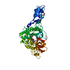
| Title | Structure of S. marcescens chitinase A, E315L, complex with hexasaccharide | |||||||||
 Components Components | chitinase A | |||||||||
 Keywords Keywords | HYDROLASE / (beta/alpha)8-barrel / oligosaccharide complex | |||||||||
| Function / homology |  Function and homology information Function and homology informationendochitinase activity / chitinase / chitin catabolic process / chitin binding / polysaccharide catabolic process Similarity search - Function | |||||||||
| Biological species |  Serratia marcescens (bacteria) Serratia marcescens (bacteria) | |||||||||
| Method |  X-RAY DIFFRACTION / SYNCHROTRON / MOLECULAR REPLACEMENT / Resolution: 2.05 Å X-RAY DIFFRACTION / SYNCHROTRON / MOLECULAR REPLACEMENT / Resolution: 2.05 Å | |||||||||
 Authors Authors | Aronson Jr., N.N. / Halloran, B.A. / Alexyev, M.F. / Amable, L. / Madura, J.D. / Pasupulati, L. / Worth, C. / Van Roey, P. | |||||||||
 Citation Citation | Journal: Biochem.J. / Year: 2003 Title: Family 18 chitinase-oligosaccharide substrate interaction: subsite preference and anomer selectivity of Serratia marcescens chitinase A. Authors: Aronson Jr., N.N. / Halloran, B.A. / Alexyev, M.F. / Amable, L. / Madura, J.D. / Pasupulati, L. / Worth, C. / Van Roey, P. | |||||||||
| History |
| |||||||||
| Remark 999 | sequence The author indicates that these represent strain variations. |
- Structure visualization
| Structure viewer | Molecule: MolmilJmol/JSmol |
|---|
- Downloads & links
-Download
| PDBx/mmCIF format | 1nh6.cif.gz | 134.7 KB | Display | PDBx/mmCIF format |
|---|---|---|---|---|
| PDB format | pdb1nh6.ent.gz | 101.4 KB | Display | PDB format |
| PDBx/mmJSON format | 1nh6.json.gz | Tree view | PDBx/mmJSON format | |
| Others |  Other downloads Other downloads |
-Validation report
| Arichive directory | https://data.pdbj.org/pub/pdb/validation_reports/nh/1nh6ftp://data.pdbj.org/pub/pdb/validation_reports/nh/1nh6 | HTTPS FTP |
|---|
-Related structure data
| Related structure data |  1edqS S: Starting model for refinement |
|---|---|
| Similar structure data |


-Links
 PDBj
PDBj- Assembly
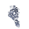
| Deposited unit | 
| ||||||||
|---|---|---|---|---|---|---|---|---|---|
| 1 |
| ||||||||
| Unit cell |
|
-Components
| #1: Protein | Mass: 58653.660 Da / Num. of mol.: 1 / Mutation: E315L Source method: isolated from a genetically manipulated source Source: (gene. exp.) Serratia marcescens (bacteria) / Plasmid: pET23a / Species (production host): Escherichia coli / Production host: References: GenBank: 3308994, UniProt: P07254*PLUS, chitinase |
|---|---|
| #2: Polysaccharide | 2-acetamido-2-deoxy-beta-D-glucopyranose-(1-4)-2-acetamido-2-deoxy-beta-D-glucopyranose-(1-4)-2- ...2-acetamido-2-deoxy-beta-D-glucopyranose-(1-4)-2-acetamido-2-deoxy-beta-D-glucopyranose-(1-4)-2-acetamido-2-deoxy-beta-D-glucopyranose-(1-4)-2-acetamido-2-deoxy-beta-D-glucopyranose-(1-4)-2-acetamido-2-deoxy-beta-D-glucopyranose-(1-4)-2-acetamido-2-deoxy-beta-D-glucopyranose Source method: isolated from a genetically manipulated source |
| #3: Water | ChemComp-HOH /  Mass: 18.015 Da / Num. of mol.: 635 / Source method: isolated from a natural source / Formula: H2O Mass: 18.015 Da / Num. of mol.: 635 / Source method: isolated from a natural source / Formula: H2O |
| Has protein modification | Y |
-Experimental details
-Experiment
| Experiment | Method: X-RAY DIFFRACTION / Number of used crystals: 1 |
|---|
- Sample preparation
| Crystal | Density Matthews: 3.65 Å3/Da / Density % sol: 66.04 % | |||||||||||||||||||||||||||||||||||||||||||||||||
|---|---|---|---|---|---|---|---|---|---|---|---|---|---|---|---|---|---|---|---|---|---|---|---|---|---|---|---|---|---|---|---|---|---|---|---|---|---|---|---|---|---|---|---|---|---|---|---|---|---|---|
| Crystal grow | Temperature: 283 K / Method: vapor diffusion, hanging drop / pH: 7 Details: 8-12% ethanol, 0.8-1.3 M NaCl, 0.1 M HEPES, pH 7.0, VAPOR DIFFUSION, HANGING DROP, temperature 283K | |||||||||||||||||||||||||||||||||||||||||||||||||
| Crystal grow | *PLUS pH: 6.5 | |||||||||||||||||||||||||||||||||||||||||||||||||
| Components of the solutions | *PLUS
|
-Data collection
| Diffraction | Mean temperature: 100 K |
|---|---|
| Diffraction source | Source: SYNCHROTRON / Site: SSRL  / Beamline: BL9-1 / Wavelength: 1 Å / Beamline: BL9-1 / Wavelength: 1 Å |
| Detector | Type: MARRESEARCH / Detector: IMAGE PLATE / Date: May 12, 2000 |
| Radiation | Protocol: SINGLE WAVELENGTH / Monochromatic (M) / Laue (L): M / Scattering type: x-ray |
| Radiation wavelength | Wavelength: 1 Å / Relative weight: 1 |
| Reflection | Resolution: 2.05→30 Å / Num. all: 681751 / Num. obs: 681751 / Observed criterion σ(I): 0 / Redundancy: 4.4 % / Biso Wilson estimate: 13.4 Å2 / Rmerge(I) obs: 0.069 / Net I/σ(I): 9.2 |
| Reflection shell | Resolution: 2.05→2.1 Å / Redundancy: 4 % / Rmerge(I) obs: 0.318 / Mean I/σ(I) obs: 2.4 / % possible all: 87.3 |
| Reflection | *PLUS Lowest resolution: 36 Å / % possible obs: 98.6 % |
| Reflection shell | *PLUS % possible obs: 87.3 % |
- Processing
| Software |
| ||||||||||||||||||||||||||||||||||||
|---|---|---|---|---|---|---|---|---|---|---|---|---|---|---|---|---|---|---|---|---|---|---|---|---|---|---|---|---|---|---|---|---|---|---|---|---|---|
| Refinement | Method to determine structure: MOLECULAR REPLACEMENT Starting model: 1EDQ Resolution: 2.05→29.16 Å / Rfactor Rfree error: 0.003 / Data cutoff high absF: 2735168.21 / Data cutoff high rms absF: 2735168.21 / Data cutoff low absF: 0 / Isotropic thermal model: RESTRAINED / Cross valid method: THROUGHOUT / σ(F): 0 / Stereochemistry target values: Engh & Huber
| ||||||||||||||||||||||||||||||||||||
| Solvent computation | Solvent model: FLAT MODEL / Bsol: 57.1079 Å2 / ksol: 0.349994 e/Å3 | ||||||||||||||||||||||||||||||||||||
| Displacement parameters | Biso mean: 25.8 Å2
| ||||||||||||||||||||||||||||||||||||
| Refine analyze |
| ||||||||||||||||||||||||||||||||||||
| Refinement step | Cycle: LAST / Resolution: 2.05→29.16 Å
| ||||||||||||||||||||||||||||||||||||
| Refine LS restraints |
| ||||||||||||||||||||||||||||||||||||
| LS refinement shell | Resolution: 2.05→2.18 Å / Rfactor Rfree error: 0.009 / Total num. of bins used: 6
| ||||||||||||||||||||||||||||||||||||
| Xplor file |
| ||||||||||||||||||||||||||||||||||||
| Software | *PLUS Version: 0.9 / Classification: refinement | ||||||||||||||||||||||||||||||||||||
| Refinement | *PLUS Lowest resolution: 36 Å / Rfactor Rfree: 0.225 / Rfactor Rwork: 0.195 | ||||||||||||||||||||||||||||||||||||
| Solvent computation | *PLUS | ||||||||||||||||||||||||||||||||||||
| Displacement parameters | *PLUS | ||||||||||||||||||||||||||||||||||||
| Refine LS restraints | *PLUS
|
