 Movie
Movie Controller

[English] 日本語
 Yorodumi
- PDB-1dxs: Crystal structure of the C-terminal sterile alpha motif (SAM) dom... -
+ Open data

- Basic information
| Entry | Database: PDB / ID: 1dxs | ||||||
|---|---|---|---|---|---|---|---|
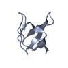
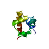
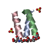
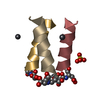
| Title | Crystal structure of the C-terminal sterile alpha motif (SAM) domain of human p73 alpha splice variant | ||||||
 Components Components | P53-LIKE TRANSCRIPTION FACTOR | ||||||
 Keywords Keywords |  GENE REGULATION / P73 SAM-LIKE DOMAIN / P53 P63 HOMOLOGUE / STERILE ALPHA MOTIF / TUMOUR SUPRESSOR GENE REGULATION / P73 SAM-LIKE DOMAIN / P53 P63 HOMOLOGUE / STERILE ALPHA MOTIF / TUMOUR SUPRESSOR | ||||||
| Function / homology |  Function and homology information Function and homology informationpositive regulation of lung ciliated cell differentiation / negative regulation of cardiac muscle cell proliferation / TP53 Regulates Transcription of Death Receptors and Ligands / Activation of PUMA and translocation to mitochondria / Regulation of TP53 Activity through Association with Co-factors / positive regulation of oligodendrocyte differentiation / TP53 regulates transcription of several additional cell death genes whose specific roles in p53-dependent apoptosis remain uncertain / TP53 Regulates Transcription of Caspase Activators and Caspases / TP53 Regulates Transcription of Genes Involved in Cytochrome C Release / negative regulation of neuron differentiation ...positive regulation of lung ciliated cell differentiation / negative regulation of cardiac muscle cell proliferation / TP53 Regulates Transcription of Death Receptors and Ligands / Activation of PUMA and translocation to mitochondria / Regulation of TP53 Activity through Association with Co-factors / positive regulation of oligodendrocyte differentiation / TP53 regulates transcription of several additional cell death genes whose specific roles in p53-dependent apoptosis remain uncertain / TP53 Regulates Transcription of Caspase Activators and Caspases / TP53 Regulates Transcription of Genes Involved in Cytochrome C Release / negative regulation of neuron differentiation / mismatch repair / intrinsic apoptotic signaling pathway in response to DNA damage by p53 class mediator / MDM2/MDM4 family protein binding / response to organonitrogen compound / regulation of mitotic cell cycle / transcription corepressor binding / kidney development / protein tetramerization / intrinsic apoptotic signaling pathway in response to DNA damage / p53 binding / cell junction / RUNX1 regulates transcription of genes involved in differentiation of HSCs / regulation of gene expression / DNA-binding transcription activator activity, RNA polymerase II-specific / DNA-binding transcription factor binding / RNA polymerase II-specific DNA-binding transcription factor binding / positive regulation of MAPK cascade / transcription cis-regulatory region binding / regulation of cell cycle / DNA-binding transcription factor activity, RNA polymerase II-specific / response to xenobiotic stimulus / cell cycle / positive regulation of apoptotic process / RNA polymerase II cis-regulatory region sequence-specific DNA binding / DNA-binding transcription factor activity / negative regulation of cell population proliferation / intracellular membrane-bounded organelle / DNA damage response / chromatin / regulation of transcription by RNA polymerase II / protein kinase binding / positive regulation of DNA-templated transcription / Golgi apparatus / positive regulation of transcription by RNA polymerase II / nucleoplasm / identical protein binding / metal ion binding / nucleus / cytosolSimilarity search - Function | ||||||
| Biological species |  HOMO SAPIENS (human) HOMO SAPIENS (human) | ||||||
| Method | X-RAY DIFFRACTION / MOLECULAR REPLACEMENT / Resolution: 2.54 Å | ||||||
 Authors Authors | Wang, W.K. / Chen, Y.W. | ||||||
 Citation Citation | Journal: Acta Crystallogr.,Sect.D / Year: 2001 Title: Structure of the C-Terminal Sterile Alpha-Motif (Sam) Domain of Human P73 Alpha Authors: Wang, W.K. / Bycroft, M. / Foster, N.W. / Buckle, A.M. / Fersht, A.R. / Chen, Y.W. #1: Journal: Acta Crystallogr.,Sect.D / Year: 2000 Title: Crystallization and Preliminary Crystallographic Studies of a Sam Domain at the C-Terminus of Human P73 Alpha Authors: Wang, W.K. / Proctor, M. / Buckle, A.M. / Bycroft, M. / Chen, Y.W. #2: Journal: Embo J. / Year: 1999Title: Solution Structure of a Conserved C-Terminal Domain of P73 with Structural Homology to the Sam Domain Authors: Chi, S.-W. / Ayed, A. / Arrowsmith, C.H. | ||||||
| History |
|
- Structure visualization
| Structure viewer | Molecule: MolmilJmol/JSmol |
|---|
- Downloads & links
-Download
| PDBx/mmCIF format | 1dxs.cif.gz | 23.7 KB | Display | PDBx/mmCIF format |
|---|---|---|---|---|
| PDB format | pdb1dxs.ent.gz | 14.3 KB | Display | PDB format |
| PDBx/mmJSON format | 1dxs.json.gz | Tree view | PDBx/mmJSON format | |
| Others |  Other downloads Other downloads |
-Validation report
| Arichive directory | https://data.pdbj.org/pub/pdb/validation_reports/dx/1dxsftp://data.pdbj.org/pub/pdb/validation_reports/dx/1dxs | HTTPS FTP |
|---|
-Related structure data
| Related structure data |  1cokS S: Starting model for refinement |
|---|---|
| Similar structure data |


-Links
 PDBj
PDBj


- Assembly
| Deposited unit | 
| |||||||||
|---|---|---|---|---|---|---|---|---|---|---|
| 1 |
| |||||||||
| Unit cell |
| |||||||||
| Components on special symmetry positions |
| |||||||||
| Details | BIOLOGICAL_UNIT: MONOMER |
-Components
| #1: Protein | Mass: 9026.124 Da / Num. of mol.: 1 / Fragment: C-TERMINAL STERILE ALPHA MOTIF (SAM) DOMAIN Source method: isolated from a genetically manipulated source Source: (gene. exp.) HOMO SAPIENS (human) / Cellular location: NUCLEUSCell nucleus / Gene: P73 / Plasmid: MODIFIED PRESET A / Cellular location (production host): CYTOPLASM / Production host:  ESCHERICHIA COLI BL21(DE3) (bacteria) / Variant (production host): C41 / References: UniProt: O15350 ESCHERICHIA COLI BL21(DE3) (bacteria) / Variant (production host): C41 / References: UniProt: O15350 |
|---|---|
| #2: Water | ChemComp-HOH / Water Mass: 18.015 Da / Num. of mol.: 26 / Source method: isolated from a natural source / Formula: H2O Mass: 18.015 Da / Num. of mol.: 26 / Source method: isolated from a natural source / Formula: H2O |
| Sequence details | RESIDUES GLY-SER (-2 TO -1) CLONING ARTIFACT |
-Experimental details
-Experiment
| Experiment | Method: X-RAY DIFFRACTION / Number of used crystals: 2 |
|---|
- Sample preparation
| Crystal | Density Matthews: 1.93 Å3/Da / Density % sol: 36 % | ||||||||||||||||||||
|---|---|---|---|---|---|---|---|---|---|---|---|---|---|---|---|---|---|---|---|---|---|
| Crystal grow | Temperature: 290 K / Method: vapor diffusion, hanging drop / pH: 8.5 Details: CRYSTALLIZED IN 0.1M TRIS-HCL PH8.5, 2.0M MONO-AMMONIUM DIHYDROGEN PHOSPHATE, AT 290K., pH 8.50 | ||||||||||||||||||||
| Crystal grow | *PLUS Temperature: 290 K / Method: vapor diffusion, hanging dropDetails: Wang, W.K., (2000) Acta Crystallogr.,Sect.D, 56, 769. | ||||||||||||||||||||
| Components of the solutions | *PLUS
|
-Data collection
| Diffraction | Mean temperature: 100 K |
|---|---|
| Diffraction source | Source: ROTATING ANODE / Type: RIGAKU RUH3R / Wavelength: 1.5418 |
| Detector | Type: MARRESEARCH / Detector: IMAGE PLATE / Date: Jun 15, 1999 / Details: MIRRORS |
| Radiation | Monochromator: NI FILTER / Protocol: SINGLE WAVELENGTH / Monochromatic (M) / Laue (L): M / Scattering type: x-ray |
| Radiation wavelength | Wavelength: 1.5418 Å / Relative weight: 1 |
| Reflection | Resolution: 2.54→26 Å / Num. obs: 2510 / % possible obs: 94.9 % / Observed criterion σ(I): 2 / Redundancy: 6.8 % / Biso Wilson estimate: 59.4 Å2 / Rmerge(I) obs: 0.073 / Rsym value: 0.067 / Net I/σ(I): 7.6 |
| Reflection shell | Resolution: 2.54→2.71 Å / Redundancy: 6.8 % / Rmerge(I) obs: 0.408 / Mean I/σ(I) obs: 2 / Rsym value: 0.377 / % possible all: 97.8 |
- Processing
| Software |
| ||||||||||||||||||||||||||||||||||||||||||||||||||||||||||||
|---|---|---|---|---|---|---|---|---|---|---|---|---|---|---|---|---|---|---|---|---|---|---|---|---|---|---|---|---|---|---|---|---|---|---|---|---|---|---|---|---|---|---|---|---|---|---|---|---|---|---|---|---|---|---|---|---|---|---|---|---|---|
| Refinement | Method to determine structure: MOLECULAR REPLACEMENT Starting model: PDB ENTRY 1COK Resolution: 2.54→26 Å / Isotropic thermal model: UNRESTRAINED / Cross valid method: THROUGHOUT / σ(F): 0 Details: C TERMINAL RESIDUES ARE NOT SEEN IN THE DENSITY MAPS
| ||||||||||||||||||||||||||||||||||||||||||||||||||||||||||||
| Solvent computation | Solvent model: MASKED FLAT MODEL / Bsol: 92.7 Å2 / ksol: 0.352 e/Å3 | ||||||||||||||||||||||||||||||||||||||||||||||||||||||||||||
| Displacement parameters | Biso mean: 62.1 Å2
| ||||||||||||||||||||||||||||||||||||||||||||||||||||||||||||
| Refine analyze |
| ||||||||||||||||||||||||||||||||||||||||||||||||||||||||||||
| Refinement step | Cycle: LAST / Resolution: 2.54→26 Å
| ||||||||||||||||||||||||||||||||||||||||||||||||||||||||||||
| Refine LS restraints |
| ||||||||||||||||||||||||||||||||||||||||||||||||||||||||||||
| LS refinement shell | Resolution: 2.54→2.63 Å / Total num. of bins used: 10
| ||||||||||||||||||||||||||||||||||||||||||||||||||||||||||||
| Xplor file |
| ||||||||||||||||||||||||||||||||||||||||||||||||||||||||||||
| Software | *PLUS Name: CNS / Version: 0.9A / Classification: refinement | ||||||||||||||||||||||||||||||||||||||||||||||||||||||||||||
| Refine LS restraints | *PLUS
|
