 Movie
Movie Controller

[English] 日本語
 Yorodumi
- PDB-1dm3: ACETYLATED BIOSYNTHETIC THIOLASE FROM ZOOGLOEA RAMIGERA IN COMPLE... -
+ Open data

- Basic information
| Entry | Database: PDB / ID: 1dm3 | ||||||
|---|---|---|---|---|---|---|---|
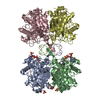
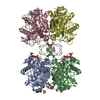
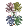
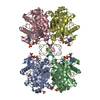
| Title | ACETYLATED BIOSYNTHETIC THIOLASE FROM ZOOGLOEA RAMIGERA IN COMPLEX WITH ACETYL-COA | ||||||
 Components Components | BIOSYNTHETIC THIOLASE ACETYLATED AT CYS89 | ||||||
 Keywords Keywords | TRANSFERASE / BIOSYNTHETIC THIOLASE / REACTION INTERMEDIATE / ACETYL-COA / BETA-ALPHA FOLD / ACETYL-COA ACETYLTRANSFERASE | ||||||
| Function / homology |  Function and homology information Function and homology informationpoly-hydroxybutyrate biosynthetic process / acetyl-CoA C-acetyltransferase / acetyl-CoA C-acetyltransferase activity / cytoplasm Similarity search - Function | ||||||
| Biological species |  Zoogloea ramigera (bacteria) Zoogloea ramigera (bacteria) | ||||||
| Method |  X-RAY DIFFRACTION / SYNCHROTRON / Resolution: 2 Å X-RAY DIFFRACTION / SYNCHROTRON / Resolution: 2 Å | ||||||
 Authors Authors | Modis, Y. / Wierenga, R.K. | ||||||
 Citation Citation | Journal: J.Mol.Biol. / Year: 2000 Title: Crystallographic analysis of the reaction pathway of Zoogloea ramigera biosynthetic thiolase. Authors: Modis, Y. / Wierenga, R.K. #1: Journal: Structure / Year: 1999Title: A biosynthetic thiolase in complex with a reaction intermediate: the crystal structure provides new insights into the catalytic mechanism Authors: Modis, Y. / Wierenga, R.K. #2: Journal: Biochemistry / Year: 1989Title: Mechanistic studies on beta-ketoacyl thiolase from Zoogloea ramigera: identification of the active-site nucleophile as Cys89, its mutation to Ser89, and kinetic and thermodynamic ...Title: Mechanistic studies on beta-ketoacyl thiolase from Zoogloea ramigera: identification of the active-site nucleophile as Cys89, its mutation to Ser89, and kinetic and thermodynamic characterization of wild-type and mutant enzymes Authors: Thompson, S. / Mayerl, F. / Peoples, O.P. / Masamune, S. / Sinskey, A.J. / Walsh, C.T. | ||||||
| History |
|
- Structure visualization
| Structure viewer | Molecule: MolmilJmol/JSmol |
|---|
- Downloads & links
-Download
| PDBx/mmCIF format | 1dm3.cif.gz | 312.8 KB | Display | PDBx/mmCIF format |
|---|---|---|---|---|
| PDB format | pdb1dm3.ent.gz | 251.2 KB | Display | PDB format |
| PDBx/mmJSON format | 1dm3.json.gz | Tree view | PDBx/mmJSON format | |
| Others |  Other downloads Other downloads |
-Validation report
| Arichive directory | https://data.pdbj.org/pub/pdb/validation_reports/dm/1dm3ftp://data.pdbj.org/pub/pdb/validation_reports/dm/1dm3 | HTTPS FTP |
|---|
-Related structure data


-Links
 PDBj
PDBj

- Assembly
| Deposited unit | 
| ||||||||
|---|---|---|---|---|---|---|---|---|---|
| 1 |
| ||||||||
| Unit cell |
|
-Components
| #1: Protein | Mass: 40297.949 Da / Num. of mol.: 4 / Fragment: ENTIRE THIOLASE, ACETYLATED AT CYS89 Source method: isolated from a genetically manipulated source Source: (gene. exp.) Zoogloea ramigera (bacteria) / Cellular location: CYTOPLASM / Plasmid: PTRC99A / Production host: #2: Chemical | ChemComp-SO4 /   Mass: 96.063 Da / Num. of mol.: 6 Mass: 96.063 Da / Num. of mol.: 6Fragment: ACETYL-COA CHAINS E,F,G AND H ARE NON-COVALENTLY BOUND TO PROTEIN CHAINS A,B,C AND D, RESPECTIVELY Source method: obtained synthetically / Formula: SO4 #3: Chemical | ChemComp-ACO /   Mass: 809.571 Da / Num. of mol.: 4 / Source method: obtained synthetically / Formula: C23H38N7O17P3S Mass: 809.571 Da / Num. of mol.: 4 / Source method: obtained synthetically / Formula: C23H38N7O17P3S#4: Water | ChemComp-HOH / |  Mass: 18.015 Da / Num. of mol.: 804 / Source method: isolated from a natural source / Formula: H2O Mass: 18.015 Da / Num. of mol.: 804 / Source method: isolated from a natural source / Formula: H2OHas protein modification | Y | |
|---|
-Experimental details
-Experiment
| Experiment | Method: X-RAY DIFFRACTION / Number of used crystals: 1 |
|---|
- Sample preparation
| Crystal | Density Matthews: 3.08 Å3/Da / Density % sol: 60.11 % | |||||||||||||||||||||||||||||||||||
|---|---|---|---|---|---|---|---|---|---|---|---|---|---|---|---|---|---|---|---|---|---|---|---|---|---|---|---|---|---|---|---|---|---|---|---|---|
| Crystal grow | Temperature: 294 K / Method: vapor diffusion, hanging drop / pH: 5 Details: 0.9 M Ammonium sulfate, 1 M lithium sulfate, 0,1 M sodium acetate, 1 mM DTT, 1mM EDTA, 1mM sodium azide, pH 5, VAPOR DIFFUSION, HANGING DROP, temperature 294K | |||||||||||||||||||||||||||||||||||
| Crystal grow | *PLUS pH: 5 | |||||||||||||||||||||||||||||||||||
| Components of the solutions | *PLUS
|
-Data collection
| Diffraction | Mean temperature: 100 K |
|---|---|
| Diffraction source | Source: SYNCHROTRON / Site: EMBL/DESY, HAMBURG  / Beamline: X31 / Wavelength: 1.057486 / Beamline: X31 / Wavelength: 1.057486 |
| Detector | Type: MARRESEARCH / Detector: AREA DETECTOR / Date: Jun 30, 1999 |
| Radiation | Protocol: SINGLE WAVELENGTH / Monochromatic (M) / Laue (L): M / Scattering type: x-ray |
| Radiation wavelength | Wavelength: 1.057486 Å / Relative weight: 1 |
| Reflection | Resolution: 2→50 Å / Num. all: 132107 / Num. obs: 126823 / % possible obs: 96 % / Observed criterion σ(F): 0 / Observed criterion σ(I): 2 / Redundancy: 2 % / Biso Wilson estimate: 25.1 Å2 / Rmerge(I) obs: 0.068 / Net I/σ(I): 10.3 |
| Reflection shell | Resolution: 2.03→2.15 Å / Redundancy: 2 % / Rmerge(I) obs: 0.22 / Num. unique all: 16710 / % possible all: 94.5 |
| Reflection | *PLUS Lowest resolution: 50 Å / % possible obs: 96 % |
| Reflection shell | *PLUS % possible obs: 94.5 % / Rmerge(I) obs: 0.22 |
- Processing
| Software |
| |||||||||||||||||||||||||
|---|---|---|---|---|---|---|---|---|---|---|---|---|---|---|---|---|---|---|---|---|---|---|---|---|---|---|
| Refinement | Resolution: 2→50 Å / σ(F): 0 / σ(I): 0 / Stereochemistry target values: Engh & Huber / Details: Weighted sparse matrix least squares procedure
| |||||||||||||||||||||||||
| Refinement step | Cycle: LAST / Resolution: 2→50 Å
| |||||||||||||||||||||||||
| Refine LS restraints |
| |||||||||||||||||||||||||
| Software | *PLUS Name: REFMAC / Classification: refinement | |||||||||||||||||||||||||
| Refinement | *PLUS Lowest resolution: 50 Å / σ(F): 0 / Rfactor obs: 0.214 | |||||||||||||||||||||||||
| Solvent computation | *PLUS | |||||||||||||||||||||||||
| Displacement parameters | *PLUS |
