 Movie
Movie Controller

[English] 日本語
 Yorodumi
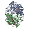
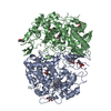
- PDB-1ddx: CRYSTAL STRUCTURE OF A MIXTURE OF ARACHIDONIC ACID AND PROSTAGLAN... -
+ Open data

- Basic information
| Entry | Database: PDB / ID: 1ddx | |||||||||
|---|---|---|---|---|---|---|---|---|---|---|
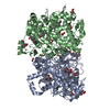
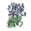
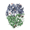
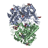
| Title | CRYSTAL STRUCTURE OF A MIXTURE OF ARACHIDONIC ACID AND PROSTAGLANDIN BOUND TO THE CYCLOOXYGENASE ACTIVE SITE OF COX-2: PROSTAGLANDIN STRUCTURE | |||||||||
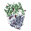
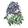
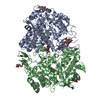
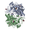
 Components Components | PROTEIN (PROSTAGLANDIN H2 SYNTHASE-2) | |||||||||
 Keywords Keywords | OXIDOREDUCTASE / COX-2 / CYCLOOXYGENASE / PROSTAGLANDIN / ARACHIDONATE / ENDOPEROXIDE | |||||||||
| Function / homology |  Function and homology information Function and homology informationBiosynthesis of DHA-derived SPMs / Biosynthesis of EPA-derived SPMs / Biosynthesis of DPAn-3 SPMs / Biosynthesis of electrophilic ω-3 PUFA oxo-derivatives / Synthesis of 15-eicosatetraenoic acid derivatives / cellular response to non-ionic osmotic stress / : / Synthesis of Prostaglandins (PG) and Thromboxanes (TX) / prostaglandin-endoperoxide synthase / prostaglandin-endoperoxide synthase activity ...Biosynthesis of DHA-derived SPMs / Biosynthesis of EPA-derived SPMs / Biosynthesis of DPAn-3 SPMs / Biosynthesis of electrophilic ω-3 PUFA oxo-derivatives / Synthesis of 15-eicosatetraenoic acid derivatives / cellular response to non-ionic osmotic stress / : / Synthesis of Prostaglandins (PG) and Thromboxanes (TX) / prostaglandin-endoperoxide synthase / prostaglandin-endoperoxide synthase activity / cyclooxygenase pathway / negative regulation of intrinsic apoptotic signaling pathway in response to osmotic stress / positive regulation of prostaglandin biosynthetic process / regulation of neuroinflammatory response / positive regulation of fever generation / response to selenium ion / prostaglandin secretion / cellular response to fluid shear stress / response to nematode / prostaglandin biosynthetic process / nuclear inner membrane / dioxygenase activity / bone mineralization / decidualization / nuclear outer membrane / keratinocyte differentiation / positive regulation of brown fat cell differentiation / brown fat cell differentiation / embryo implantation / peroxidase activity / regulation of blood pressure / regulation of cell population proliferation / response to oxidative stress / cellular response to hypoxia / neuron projection / heme binding / endoplasmic reticulum membrane / negative regulation of apoptotic process / enzyme binding / protein homodimerization activity / metal ion binding / cytoplasm / cytosol Similarity search - Function | |||||||||
| Biological species |  | |||||||||
| Method |  X-RAY DIFFRACTION / Resolution: 3 Å X-RAY DIFFRACTION / Resolution: 3 Å | |||||||||
 Authors Authors | Kiefer, J.R. / Pawlitz, J.L. / Moreland, K.T. / Stegeman, R.A. / Gierse, J.K. / Stevens, A.M. / Goodwin, D.C. / Rowlinson, S.W. / Marnett, L.J. / Stallings, W.C. / Kurumbail, R.G. | |||||||||
 Citation Citation | Journal: Nature / Year: 2000 Title: Structural insights into the stereochemistry of the cyclooxygenase reaction. Authors: Kiefer, J.R. / Pawlitz, J.L. / Moreland, K.T. / Stegeman, R.A. / Hood, W.F. / Gierse, J.K. / Stevens, A.M. / Goodwin, D.C. / Rowlinson, S.W. / Marnett, L.J. / Stallings, W.C. / Kurumbail, R.G. #1: Journal: Nature / Year: 1996Title: Structural Basis for Selective Inhibition of Cyclooxygenase-2 by Anti- Inflammatory Agents Authors: Kurumbail, R.G. / Stevens, A.M. / Gierse, J.K. / McDonald, J.J. / Stegeman, R.A. / Pak, J.Y. / Gildehaus, D. / Miyashiro, J.M. / Penning, T.D. / Seibert, K. / Isakson, P.C. / Stallings, W.C. | |||||||||
| History |
|
- Structure visualization
| Structure viewer | Molecule: MolmilJmol/JSmol |
|---|
- Downloads & links
-Download
| PDBx/mmCIF format | 1ddx.cif.gz | 464.2 KB | Display | PDBx/mmCIF format |
|---|---|---|---|---|
| PDB format | pdb1ddx.ent.gz | 371.7 KB | Display | PDB format |
| PDBx/mmJSON format | 1ddx.json.gz | Tree view | PDBx/mmJSON format | |
| Others |  Other downloads Other downloads |
-Validation report
| Arichive directory | https://data.pdbj.org/pub/pdb/validation_reports/dd/1ddxftp://data.pdbj.org/pub/pdb/validation_reports/dd/1ddx | HTTPS FTP |
|---|
-Related structure data


-Links
 PDBj
PDBj


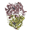
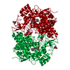
- Assembly
| Deposited unit | 
| ||||||||
|---|---|---|---|---|---|---|---|---|---|
| 1 | 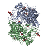
| ||||||||
| 2 | 
| ||||||||
| Unit cell |
|
-Components
-Protein , 1 types, 4 molecules ABCD
| #1: Protein | Mass: 63497.422 Da / Num. of mol.: 4 Source method: isolated from a genetically manipulated source Source: (gene. exp.)   Spodoptera frugiperda (fall armyworm) Spodoptera frugiperda (fall armyworm)References: UniProt: Q05769, prostaglandin-endoperoxide synthase |
|---|
-Sugars , 3 types, 16 molecules 

| #2: Polysaccharide | 2-acetamido-2-deoxy-beta-D-glucopyranose-(1-4)-2-acetamido-2-deoxy-beta-D-glucopyranose Source method: isolated from a genetically manipulated source #3: Sugar | ChemComp-NAG /  Type: D-saccharide, beta linking / Mass: 221.208 Da / Num. of mol.: 8 Type: D-saccharide, beta linking / Mass: 221.208 Da / Num. of mol.: 8Source method: isolated from a genetically manipulated source Formula: C8H15NO6 #4: Sugar | ChemComp-BOG /  Type: D-saccharide / Mass: 292.369 Da / Num. of mol.: 4 / Source method: obtained synthetically / Formula: C14H28O6 / Comment: detergent*YM Type: D-saccharide / Mass: 292.369 Da / Num. of mol.: 4 / Source method: obtained synthetically / Formula: C14H28O6 / Comment: detergent*YM |
|---|
-Non-polymers , 2 types, 177 molecules 

| #5: Chemical | ChemComp-PGX /  Mass: 368.464 Da / Num. of mol.: 4 / Source method: obtained synthetically / Formula: C20H32O6 Mass: 368.464 Da / Num. of mol.: 4 / Source method: obtained synthetically / Formula: C20H32O6#6: Water | ChemComp-HOH / | Mass: 18.015 Da / Num. of mol.: 173 / Source method: isolated from a natural source / Formula: H2O |
|---|
-Details
| Has protein modification | Y |
|---|
-Experimental details
-Experiment
| Experiment | Method: X-RAY DIFFRACTION / Number of used crystals: 1 |
|---|
- Sample preparation
| Crystal | Density Matthews: 2.93 Å3/Da / Density % sol: 57.99 % | ||||||||||||||||||||||||||||||||||||||||||||||||||||||||||||||||||||||||||||||||||||
|---|---|---|---|---|---|---|---|---|---|---|---|---|---|---|---|---|---|---|---|---|---|---|---|---|---|---|---|---|---|---|---|---|---|---|---|---|---|---|---|---|---|---|---|---|---|---|---|---|---|---|---|---|---|---|---|---|---|---|---|---|---|---|---|---|---|---|---|---|---|---|---|---|---|---|---|---|---|---|---|---|---|---|---|---|---|
| Crystal grow | Temperature: 288 K / Method: vapor diffusion, hanging drop / pH: 8 Details: MONOMETHYL PEG 550, MAGNESIUM SULFATE, ARACHIDNOIC ACID, EPPS, B-OG, pH 8.00, VAPOR DIFFUSION, HANGING DROP, temperature 288K | ||||||||||||||||||||||||||||||||||||||||||||||||||||||||||||||||||||||||||||||||||||
| Crystal grow | *PLUS Temperature: 18 ℃ / Details: Stevens, A.M., (1999) J. Cryst. Growth., 196, 350. / pH: 8 | ||||||||||||||||||||||||||||||||||||||||||||||||||||||||||||||||||||||||||||||||||||
| Components of the solutions | *PLUS
|
-Data collection
| Diffraction | Mean temperature: 130 K |
|---|---|
| Diffraction source | Source: ROTATING ANODE / Type: RIGAKU RU300 / Wavelength: 1.514 |
| Detector | Type: MARRESEARCH / Detector: IMAGE PLATE / Date: Aug 7, 1997 / Details: DOUBLE-MIRROR OPTICS |
| Radiation | Protocol: SINGLE WAVELENGTH / Monochromatic (M) / Laue (L): M / Scattering type: x-ray |
| Radiation wavelength | Wavelength: 1.514 Å / Relative weight: 1 |
| Reflection | Resolution: 3→40 Å / Num. obs: 45048 / % possible obs: 96.2 % / Observed criterion σ(I): -3 / Redundancy: 3.4 % / Biso Wilson estimate: 16.9 Å2 / Rmerge(I) obs: 0.112 / Rsym value: 0.112 / Net I/σ(I): 3 |
| Reflection | *PLUS Highest resolution: 3 Å / Lowest resolution: 40 Å / Observed criterion σ(I): -3 / Redundancy: 3.4 % |
- Processing
| Software |
| ||||||||||||||||||||||||||||||||||||||||
|---|---|---|---|---|---|---|---|---|---|---|---|---|---|---|---|---|---|---|---|---|---|---|---|---|---|---|---|---|---|---|---|---|---|---|---|---|---|---|---|---|---|
| Refinement | Resolution: 3→20 Å / Rfactor Rfree error: 0.005 / Data cutoff high absF: 10000000 / Data cutoff low absF: 0.001 / Isotropic thermal model: RESTRAINED / Cross valid method: THROUGHOUT / σ(F): 0 / σ(I): -1 / Stereochemistry target values: Engh & Huber
| ||||||||||||||||||||||||||||||||||||||||
| Displacement parameters | Biso mean: 28.3 Å2
| ||||||||||||||||||||||||||||||||||||||||
| Refine analyze |
| ||||||||||||||||||||||||||||||||||||||||
| Refinement step | Cycle: LAST / Resolution: 3→20 Å
| ||||||||||||||||||||||||||||||||||||||||
| Refine LS restraints |
| ||||||||||||||||||||||||||||||||||||||||
| LS refinement shell | Resolution: 3→3.19 Å / Rfactor Rfree error: 0.019 / Total num. of bins used: 6
| ||||||||||||||||||||||||||||||||||||||||
| Xplor file |
| ||||||||||||||||||||||||||||||||||||||||
| Software | *PLUS Name: X-PLOR / Version: 3.851 / Classification: refinement | ||||||||||||||||||||||||||||||||||||||||
| Refinement | *PLUS σ(F): 0 / % reflection Rfree: 9.4 % | ||||||||||||||||||||||||||||||||||||||||
| Solvent computation | *PLUS | ||||||||||||||||||||||||||||||||||||||||
| Displacement parameters | *PLUS Biso mean: 28.3 Å2 | ||||||||||||||||||||||||||||||||||||||||
| Refine LS restraints | *PLUS
| ||||||||||||||||||||||||||||||||||||||||
| LS refinement shell | *PLUS Rfactor Rfree: 0.447 / % reflection Rfree: 9.5 % / Rfactor Rwork: 0.356 |
