 Movie
Movie Controller

[English] 日本語
 Yorodumi
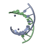
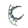
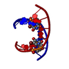
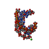
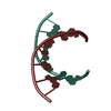
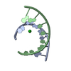
- PDB-1d92: REFINED CRYSTAL STRUCTURE OF AN OCTANUCLEOTIDE DUPLEX WITH G.T MI... -
+ Open data

- Basic information
| Entry | Database: PDB / ID: 1d92 | ||||||||||||||||||
|---|---|---|---|---|---|---|---|---|---|---|---|---|---|---|---|---|---|---|---|
| Title | REFINED CRYSTAL STRUCTURE OF AN OCTANUCLEOTIDE DUPLEX WITH G.T MISMATCHED BASE-PAIRS | ||||||||||||||||||
 Components Components | DNA (5'-D(* Keywords Keywords DNA / A-DNA / DOUBLE HELIX / MISMATCHED DNA / A-DNA / DOUBLE HELIX / MISMATCHEDFunction / homology | DNA Function and homology information Function and homology informationMethod | X-RAY DIFFRACTION / Resolution: 2.25 Å  Authors AuthorsHunter, W.N. / Kneale, G. / Brown, T. / Rabinovich, D. / Kennard, O. |  CitationJournal: J.Mol.Biol. / Year: 1986 CitationJournal: J.Mol.Biol. / Year: 1986Title: Refined crystal structure of an octanucleotide duplex with G . T mismatched base-pairs. Authors: Hunter, W.N. / Kneale, G. / Brown, T. / Rabinovich, D. / Kennard, O. #1: Journal: Nature / Year: 1985Title: High-Resolution Structure of a DNA Helix Containing Mismatched Base Pairs Authors: Brown, T. / Kennard, O. / Kneale, G. / Rabinovich, D. #2: Journal: J.Biomol.Struct.Dyn. / Year: 1985Title: Structural Studies of DNA Fragments. The G.T Wobble Base Pair in A, B and Z DNA. The G.A Base Pair in B-DNA Authors: Kennard, O. History |
|
- Structure visualization
| Structure viewer | Molecule: MolmilJmol/JSmol |
|---|
- Downloads & links
-Download
| PDBx/mmCIF format | 1d92.cif.gz | 17.7 KB | Display | PDBx/mmCIF format |
|---|---|---|---|---|
| PDB format | pdb1d92.ent.gz | 11.5 KB | Display | PDB format |
| PDBx/mmJSON format | 1d92.json.gz | Tree view | PDBx/mmJSON format | |
| Others |  Other downloads Other downloads |
-Validation report
| Arichive directory | https://data.pdbj.org/pub/pdb/validation_reports/d9/1d92ftp://data.pdbj.org/pub/pdb/validation_reports/d9/1d92 | HTTPS FTP |
|---|
-Related structure data
| Similar structure data |
|---|


-Links
 PDBj
PDBj

- Assembly
| Deposited unit | 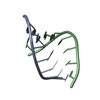
| ||||||||
|---|---|---|---|---|---|---|---|---|---|
| 1 |
| ||||||||
| Unit cell |
|
-Components
| #1: DNA chain | Mass: 2443.604 Da / Num. of mol.: 2 / Source method: obtained synthetically #2: Water | ChemComp-HOH / | Water Mass: 18.015 Da / Num. of mol.: 52 / Source method: isolated from a natural source / Formula: H2O Mass: 18.015 Da / Num. of mol.: 52 / Source method: isolated from a natural source / Formula: H2O |
|---|
-Experimental details
-Experiment
| Experiment | Method: X-RAY DIFFRACTION |
|---|
- Sample preparation
| Crystal | Density Matthews: 2.59 Å3/Da / Density % sol: 52.56 % | |||||||||||||||||||||||||
|---|---|---|---|---|---|---|---|---|---|---|---|---|---|---|---|---|---|---|---|---|---|---|---|---|---|---|
| Crystal grow | Temperature: 277 K / Method: vapor diffusion / pH: 6.5 / Details: pH 6.50, VAPOR DIFFUSION, temperature 277.00K | |||||||||||||||||||||||||
| Components of the solutions |
| |||||||||||||||||||||||||
| Crystal grow | *PLUS Temperature: 4 ℃ / pH: 6.5 / Method: other | |||||||||||||||||||||||||
| Components of the solutions | *PLUS
|
-Data collection
| Diffraction | Mean temperature: 275 K |
|---|---|
| Detector | Type: SYNTEX P21 / Detector: DIFFRACTOMETER |
| Radiation | Scattering type: x-ray |
| Radiation wavelength | Relative weight: 1 |
| Reflection | Highest resolution: 2.25 Å / Num. obs: 2414 |
| Reflection | *PLUS Highest resolution: 2.25 Å / Rmerge(I) obs: 0.06 |
- Processing
| Software | Name: NUCLSQ / Classification: refinement | ||||||||||||||||||||||||
|---|---|---|---|---|---|---|---|---|---|---|---|---|---|---|---|---|---|---|---|---|---|---|---|---|---|
| Refinement | Resolution: 2.25→10 Å / σ(I): 2 /
| ||||||||||||||||||||||||
| Refine Biso |
| ||||||||||||||||||||||||
| Refinement step | Cycle: LAST / Resolution: 2.25→10 Å
| ||||||||||||||||||||||||
| Refinement | *PLUS Highest resolution: 2.25 Å / Lowest resolution: 10 Å / Num. reflection obs: 1924 / σ(I): 2 / Rfactor obs: 0.136 | ||||||||||||||||||||||||
| Solvent computation | *PLUS | ||||||||||||||||||||||||
| Displacement parameters | *PLUS Biso mean: 85 Å2 | ||||||||||||||||||||||||
| Refine LS restraints | *PLUS
|
