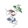
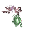
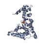
Mass: 28443.596 Da / Num. of mol.: 1 / Mutation: N-TERMINAL HIS-TAGGED Source method: isolated from a genetically manipulated source Source: (gene. exp.) Rous sarcoma virus / Genus: Alpharetrovirus / Strain: SCHMIDT RUPPIN A-2 Description: THE SOURCE OF THE CA_RSV GENE IS PLASMID PSRA-2 OF ATCC 45000, WHICH IS A PLASMID CLONE CONTAINING FULL LENGTH ROUS SARCOMA VIRUS, STRAIN SCHMIDT RUPPIN A-2, DERIVED FROM UNINTEGRATED ...Description: THE SOURCE OF THE CA_RSV GENE IS PLASMID PSRA-2 OF ATCC 45000, WHICH IS A PLASMID CLONE CONTAINING FULL LENGTH ROUS SARCOMA VIRUS, STRAIN SCHMIDT RUPPIN A-2, DERIVED FROM UNINTEGRATED DNA FROM AN ACUTE INFECTION OF QT-6 CELLS Plasmid: PET-16B (NOVAGEN) / Production host: Escherichia coli (E. coli) / Strain (production host): HMS174(DE3) PLYSS / References: UniProt: O92954
-
Experimental details
-
Experiment
Experiment
Method: SOLUTION NMR
NMR experiment
Conditions-ID
Experiment-ID
Solution-ID
Type
1
1
1
4D 13C-SEPARATED NOESY
1
2
1
4D 13C/15N-SEPARATED NOESY
1
3
1
4D 15N-SEPARATED NOESY
1
4
1
3D 15N- SEPARATED NOESY
NMR details
Text: THE 1H, 13C, AND 15N CHEMICAL SHIFT ASSIGNMENTS HAVE BEEN DEPOSITED ATTHE BIOMOLECULAR RESONANCE DATABANK (BMRB ENTRY 4384).
-
Sample preparation
Details
Solution-ID
Contents
Solvent system
1
~1 MM CA_RSV, 10 MM PHOSPHATE (PH 6.0), 0.2 MM N3NA, 0.1 MM EDTA, 0.1 MM PMSF, 1 MG/L PEPSTATIN A, 5 MM BETA-MERCAPTOETHANOL
90% H2O/10% D2O
2
~1 MM CA_RSV, 10 MM PHOSPHATE (PH 6.0), 0.2 MM N3NA, 0.1 MM EDTA, 0.1 MM PMSF, 1 MG/L PEPSTATIN A, 5 MM BETA-MERCAPTOETHANOL
100% D2O
Sample conditions
Ionic strength: 10 mM PHOSPHATE / pH: 6 / Pressure: 1 atm / Temperature: 303.00 K
Crystal grow
*PLUS
Method: other / Details: NMR
-
NMR measurement
NMR spectrometer
Type: Bruker DRX / Manufacturer: Bruker / Model: DRX / Field strength: 800 MHz
-
Processing
NMR software
Name
Version
Developer
Classification
DYANA
1.5
P.GUNTHER, ETH-ZURICH
refinement
XwinNMR
2.5
structuresolution
NMRPipe
1999
structuresolution
NMRView
3
structuresolution
TALOS
1999.019.15.47
structuresolution
Refinement
Method: TORSION ANGLE DYNAMICS SIMULATED ANNEALING / Software ordinal: 1 Details: 20 CONFORMERS COMPATIBLE WITH THE NMR CONSTRAINTS WERE CALCULATED USING DYANA 1.5 AND THE STANDARD TORSION ANGLE SIMULATED ANHEALING PROTOCOL. PRELIMINARY CALCULATIONS WERE CARRIED OUT ...Details: 20 CONFORMERS COMPATIBLE WITH THE NMR CONSTRAINTS WERE CALCULATED USING DYANA 1.5 AND THE STANDARD TORSION ANGLE SIMULATED ANHEALING PROTOCOL. PRELIMINARY CALCULATIONS WERE CARRIED OUT INDEPENDENTLY FOR EACH OF THE TWO N- AND C- TERMINAL DOMAINS OF THE PROTEIN. INITIALLY ONLY NOE DISTANCE CONSTRAINTS WERE IMPOSED. THE INITIAL STRUCTURES WERE THEN USED TO ASSES THE ACCURACY OF THE TORSION ANGLE CONSTRAINTS GENERATED BY ANALYSIS OF HA, CA, CB, CO AND N CHEMICAL SHIFTS WITH THE PROGRAM TALOS. FINALLY, UPPER AND LOWER LIMIT DISTANCE CONSTRAINTS WERE IMPOSED FOR UNAMBIGUOUS INTRAHELICAL H-BONDS.
NMR ensemble
Conformer selection criteria: target function / Conformers calculated total number: 20 / Conformers submitted total number: 20
+
About Yorodumi
-
News
-
Feb 9, 2022. New format data for meta-information of EMDB entries
New format data for meta-information of EMDB entries
Version 3 of the EMDB header file is now the official format.
The previous official version 1.9 will be removed from the archive.
In the structure databanks used in Yorodumi, some data are registered as the other names, "COVID-19 virus" and "2019-nCoV". Here are the details of the virus and the list of structure data.
Jan 31, 2019. EMDB accession codes are about to change! (news from PDBe EMDB page)
EMDB accession codes are about to change! (news from PDBe EMDB page)
The allocation of 4 digits for EMDB accession codes will soon come to an end. Whilst these codes will remain in use, new EMDB accession codes will include an additional digit and will expand incrementally as the available range of codes is exhausted. The current 4-digit format prefixed with “EMD-” (i.e. EMD-XXXX) will advance to a 5-digit format (i.e. EMD-XXXXX), and so on. It is currently estimated that the 4-digit codes will be depleted around Spring 2019, at which point the 5-digit format will come into force.
The EM Navigator/Yorodumi systems omit the EMD- prefix.
Related info.:Q: What is EMD? / ID/Accession-code notation in Yorodumi/EM Navigator
Yorodumi is a browser for structure data from EMDB, PDB, SASBDB, etc.
This page is also the successor to EM Navigator detail page, and also detail information page/front-end page for Omokage search.
The word "yorodu" (or yorozu) is an old Japanese word meaning "ten thousand". "mi" (miru) is to see.
Related info.:EMDB / PDB / SASBDB / Comparison of 3 databanks / Yorodumi Search / Aug 31, 2016. New EM Navigator & Yorodumi / Yorodumi Papers / Jmol/JSmol / Function and homology information / Changes in new EM Navigator and Yorodumi
 Movie
Movie Controller

 Yorodumi
 Open data
 Basic information
 Components
 Keywords
 Function and homology information
 Rous sarcoma virus
 Authors
 Citation
 Structure visualization
 Downloads & links
 Other downloads

 PDBj
PDBj Assembly

 Sample preparation
 Processing
 NMRPipe